

CT-guided biopsy of focal lesions in patients with multiple myeloma may reveal new and more aggressive cytogenetic abnormalities
- PMID: 11290500
- PMCID: PMC7976013
CT-guided biopsy of focal lesions in patients with multiple myeloma may reveal new and more aggressive cytogenetic abnormalities
Abstract
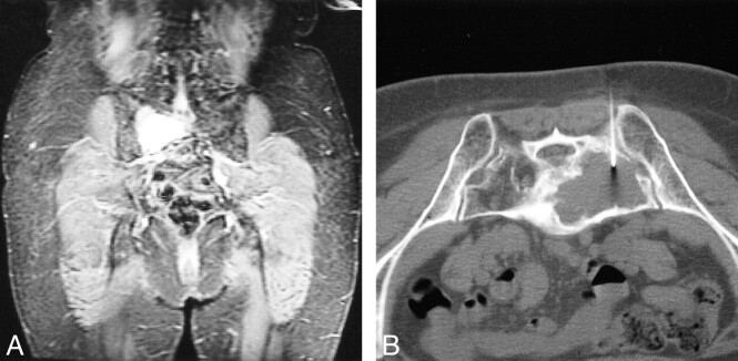
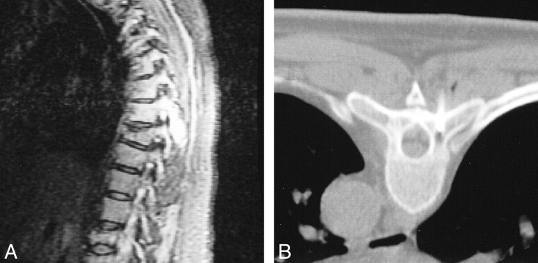
Background and purpose: Cytogenetic abnormalities, especially chromosome 13 deletion, are high-risk factors for multiple myeloma. Attaining the highest detection rates of cytogenetic abnormalities is important to provide accurate prognostic information to the referring oncologist. The purpose of this study was to use CT-guided percutaneous fine-needle aspiration bone biopsy (CT-guided FNA) of MR-detected focal lesions in patients with multiple myeloma to increase identification of abnormal cytogenetics.
Methods: Patients enrolled in two clinical trials for myeloma therapy underwent MR imaging of the entire spine and pelvis. CT-guided FNA biopsy samples obtained from MR-detected focal lesions in these patients were sent for cytogenetic analysis. FNA results were then compared with random bone marrow sampling of the iliac crest done at or near the same time as the FNA to provide the data revealed in this study.
Results: Forty-one patients (47 lesions) in one of the trials and 37 patients (38 lesions) in the other trial had biopsies performed. CT-guided FNA revealed cytogenetic abnormalities in 21% of the total patient population and new information in nearly 10% of the patients in one trial and in 20% of those in the other trial.
Conclusion: CT-guided biopsy of MR-detected focal lesions is a safe technique that can provide important cytogenetic information in a significant number of patients with multiple myeloma not identified during random marrow sampling.
Figures
References
-
- Barlogie B. Plasma cell myeloma. In: Quips T, ed. William's Hematology. 5th ed. New York: McGraw-Hill 1995;1109-1126
-
- Durie BG, Salmon SE. A clinical staging system for multiple myeloma: correlation of measured myeloma cell mass with presenting clinical features, response to treatment and survival. Cancer 1975;36:842-854 - PubMed
-
- Kyle R. Why better prognostic factors for multiple myeloma are needed. Blood 1994;83:1713-1716 - PubMed
-
- Desikan R, Barlogie B, Sawyer JR, et al. Results of high dose therapy for 1,000 patients with multiple myeloma: durable complete remissions and superior survival in the absence of chromosome 13 abnormalities. Blood 2000;95:4008-4010 - PubMed
-
- Tricot G, Sawyer JR, Jagannath S, et al. Unique role of cytogenetics in the prognosis of patients with myeloma receiving high-dose therapy and autotransplants. J Clin Oncol 1997;15:2659-2666 - PubMed
MeSH terms
LinkOut - more resources
Full Text Sources
Medical