. 2001 Apr;158(4):1271-8.
doi: 10.1016/S0002-9440(10)64078-3.

Relocalization of apoptosis-inducing factor in photoreceptor apoptosis induced by retinal detachment in vivo

Affiliations

Relocalization of apoptosis-inducing factor in photoreceptor apoptosis induced by retinal detachment in vivo

T Hisatomi et al. Am J Pathol. 2001 Apr.

Abstract

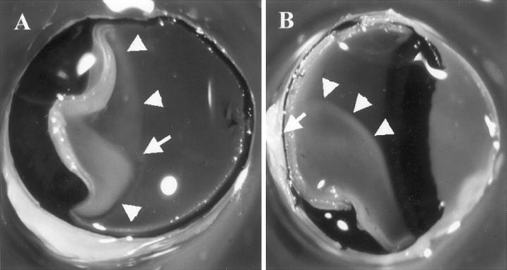
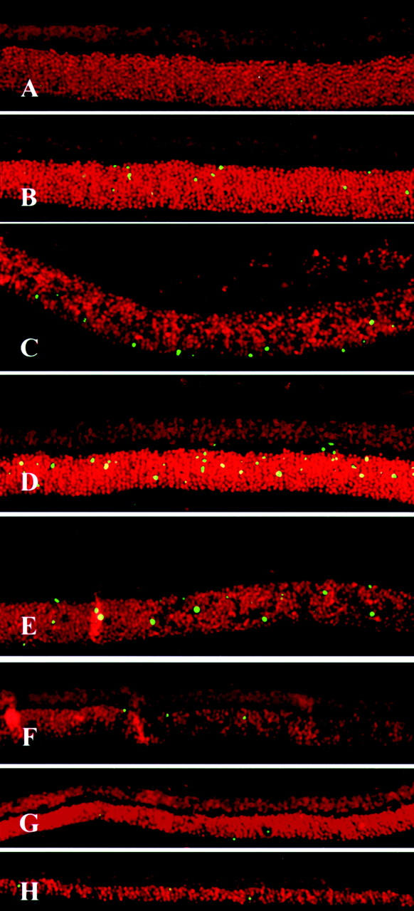
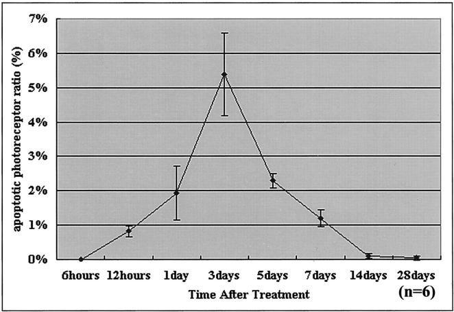
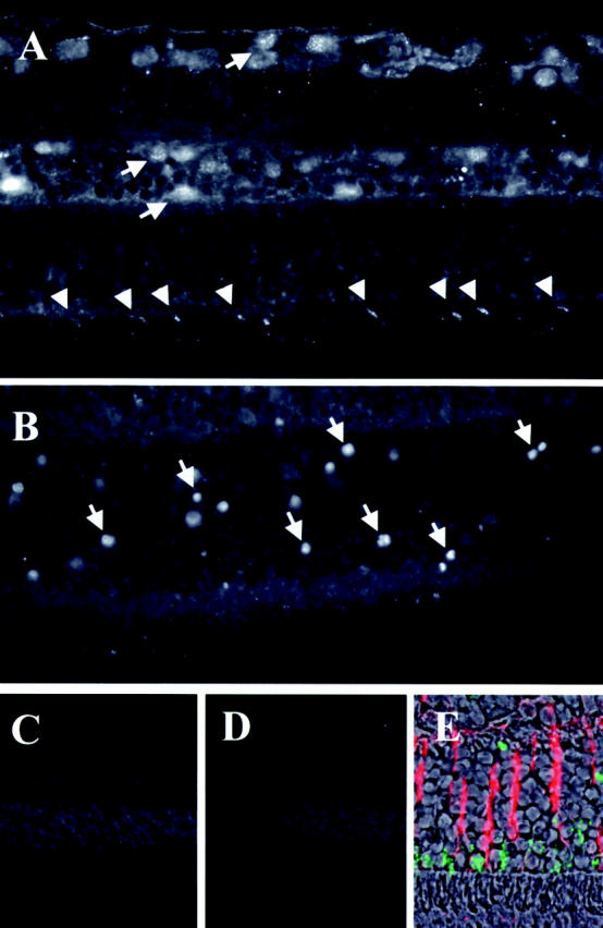
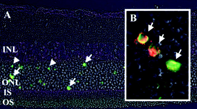
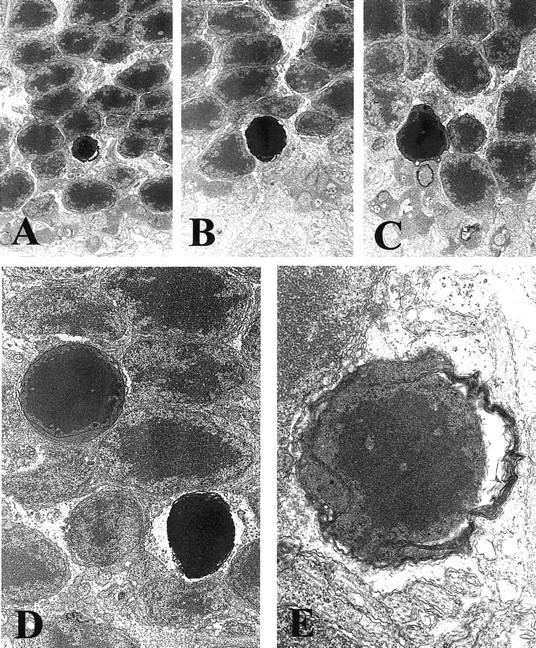
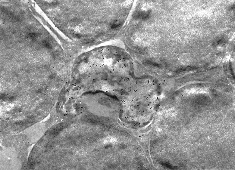
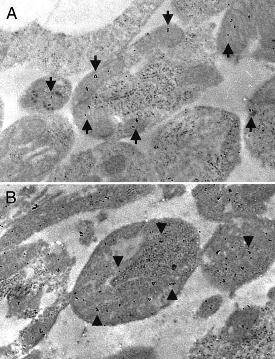
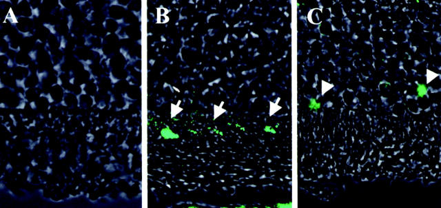
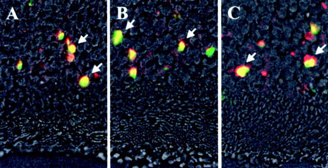
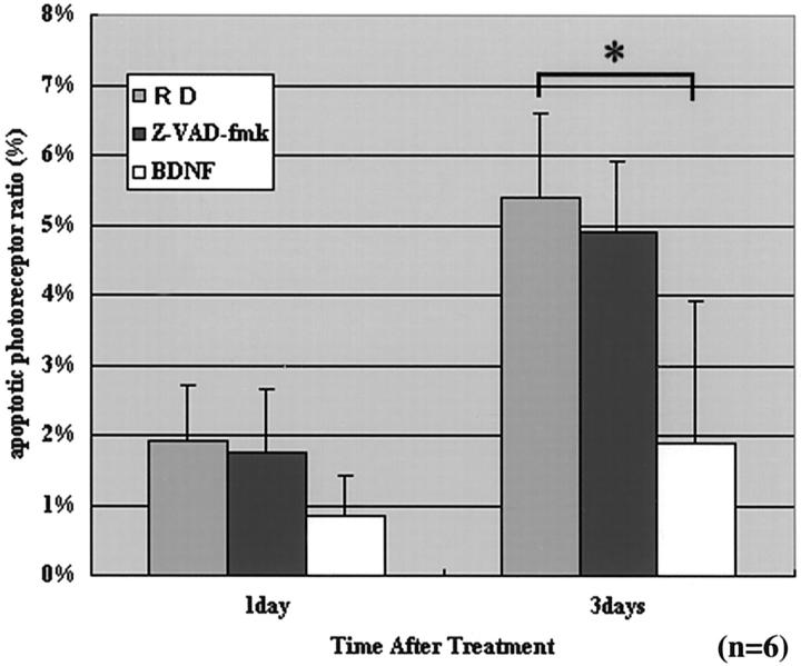
Apoptosis-inducing factor (AIF) is a novel mediator in apoptosis. AIF is a flavoprotein that is normally confined to the mitochondrial intermembrane space, yet translocates to the nucleus in several in vitro models of apoptosis. To investigate the role of AIF in the apoptotic process in vivo, we induced retinal detachment (RD) by subretinal injection of sodium hyaluronate, either in Brown Norway rats or in C3H mice. Apoptotic DNA fragmentation, as determined by terminal nick-end labeling, was most prominent 3 days after RD. The subcellular localization of AIF was examined by immunohistochemistry and immunoelectron microscopy. In normal photoreceptor cells, AIF was present in the mitochondrion-rich inner segment. However, AIF was found in the nucleus after RD. Photoreceptor apoptosis developed similarly in C3H control mice, and in mice bearing the gld or lpr mutations, indicating that cell death occurs independently from the CD95/CD95 ligand system. Both the mitochondrio-nuclear transition of AIF localization and the nuclear DNA fragmentation were inhibited by subretinal application of brain-derived neurotrophic factor. To our knowledge, this is the first description of AIF relocalization occurring in a clinically relevant, in vivo model of apoptosis.


Figures

Figure 1.
Figure 1.
Rat eyes with RD. The eyes were fixed in 1% glutaraldehyde and 1% paraformaldehyde in PBS, cut in half, and the lens removed. Note the grayish detached retina (arrowheads). The arrow indicates the optic nerve head. A: A frontal section observed from the anterior side. B: A sagittal section.
Figure 2.
Figure 2.
Fluorescent micrographs by TUNEL of sections from experimental RD from 6 hours to 28 days after treatment. Each section was stained with TdT-dUTP TUNEL and propidium iodide (see Materials and Methods). Note the TUNEL-positive cells in the photoreceptor layer (green). The highest frequency of TUNEL-positive photoreceptors is observed 3 days after treatment. The thickness of the outer nuclear layer decreases throughout time. A: 6 hours after treatment; B, 12 hours; C, 1 day, D, 3 days; E, 5 days; F, 7 days; G, 14 days; H, 28 days. Original magnifications, ×200.
Figure 3.
Figure 3.
Time course of photoreceptor apoptosis in experimental RD detected by TdT-dUTP TUNEL. The ratio of the number of apoptotic photoreceptors/total photoreceptors increases for 3 days after detachment and then gradually decreases. Apoptotic photoreceptor ratio (%) was evaluated as the ratio of total number of TUNEL-positive photoreceptors/total number of photoreceptors in each section. The sections for each eye specimen were randomly selected and observed by masked observers (six eyes for each time point). The representative results of three independent experiments are presented.
Figure 4.
Figure 4.
Fluorescent micrographs of AIF immunohistochemistry. The eyes were stained by AIF antiserum and visualized by fluorescent microscopy (see Materials and Methods). A: Control retina stained by AIF-antiserum. B: Detached retina on day 3 stained by AIF-antiserum. C: Control for staining by pre-immune serum. D: Control by pre-absorbed antiserum (with 1 μg/μl recombinant AIF). E: Double staining for AIF (green) and GFAP (red) on day 1 after RD. Specific AIF-staining is shown in ganglion cells, inner nuclear layer, outer plexiform layer, and photoreceptors (A, arrows). In normal photoreceptors, AIF staining is localized in inner segment, showing a multilinear pattern (A, arrowheads). Note the AIF-positive photoreceptor nuclei in the outer nuclear layer of detached retina (B, arrows). AIF staining decreased from the inner segment and appeared in the nucleus. Original magnifications: ×400 (A, B, and E), ×200 (C and D).
Figure 5.
Figure 5.
Fluorescent microphotographs of the detached retina, double stained by AIF and TUNEL. Each section was double stained by TUNEL (green) and with AIF antiserum (red), and a micrograph was made using a phase-contrast microscope. A: Detached retina (INL, inner nuclear layer; ONL, outer nuclear layer; IS, inner segment; OS, outer segment). Most of AIF-positive photoreceptors (arrow) are also TUNEL-positive (arrowhead), indicating apoptotic photoreceptors. B: High magnification of the outer nuclear layer. Co-localization of TUNEL and AIF staining is shown in detail. Original magnifications: ×400 (A), ×1000 (B).
Figure 6.
Figure 6.
Electron microscopic photographs of the apoptotic photoreceptors. Eyes were fixed in 1% glutaraldehyde and 1% paraformaldehyde in PBS, and embedded in Epon. Major characteristic changes occurring in apoptosis, such as chromatin condensation, alteration of nuclear shape, and cell shrinkage, are seen in the photoreceptors. Original magnification, ×10,000.
Figure 7.
Figure 7.
Immunoelectron microscopic photograph of the apoptotic photoreceptors. Apoptotic photoreceptor nuclei shown in Figure 6 ▶ are seen as irregularly shaped nuclei in the sections fixed in 1% paraformaldehyde and embedded in LR white. AIF is positive in the cytoplasm and nucleus of apoptotic photoreceptors. The dense staining of AIF on the nucleus is remarkable. Normal photoreceptors are negative in AIF staining. Original magnification, ×10,000.
Figure 8.
Figure 8.
Immunoelectron microscopic photographs of the inner segment of the photoreceptors. A: Inner segment of the photoreceptor from the control retina (nondetached). B: Inner segment of the photoreceptor from the detached retina (day 3). Mitochondria is placed regularly and the cell structure is well preserved, and AIF staining is confined to mitochondria (A, arrows). In detached retina, mitochondria degenerates and AIF staining is diffusely distributed in the cytosol of destructive cells (B, arrowheads). Original magnifications, ×13,000).
Figure 9.
Figure 9.
Fluorescent micrographs of cytochrome c immunohistochemistry. The eyes were stained by cytochrome c antibody and visualized by fluorescent microscopy. A: Control for staining without primary antibody. B: Control retina stained by cytochrome c antibody. Cytochrome c is positive in multilinear pattern in the inner segment of the photoreceptor (arrows). C: Detached retina stained by cytochrome c antibody. Cytochrome c is positive in the cytosol and nucleus in the outer nuclear layer (arrowheads).
Figure 10.
Figure 10.
Fluorescent microphotographs of double staining by AIF and TUNEL in CD95/CD-95 ligand gene mutation mice. A, C3H, control mice; B, C3H-lpr, CD95 gene mutation; C, C3H-gld, CD95 ligand gene mutation. AIF immunohistochemistry (red), TUNEL method (green), and phase-contrast microscopy on day 3 after RD. Double-positive photoreceptors (yellow, arrows) by TUNEL and AIF are seen in all groups.
Figure 11.
Figure 11.
The effect of Z-VAD.fmk and BDNF on photoreceptor apoptosis on days 1 and 3. Ten sections of each eye specimen were randomly selected and observed in a masked manner (n = 6). The wide-ranging caspase inhibitor Z-VAD.fmk does not inhibit photoreceptor apoptosis significantly, but the neurotrophic factor BDNF does (on day 3) (P < 0.05).

References

    1. Kerr JF, Wyllie AH, Currie AR: Apoptosis: a basic biological phenomenon with wide-ranging implications in tissue kinetics. Br J Cancer 1972, 26:239-257 - PMC - PubMed
    1. Huppertz B, Frank HG, Kaufmann P: The apoptosis cascade—morphological and immunohistochemical methods for its visualization. Anat Embryol (Berl) 1999, 200:1-18 - PubMed
    1. Kroemer G, Dallaporta B, Resche-Rigon M: The mitochondrial death/life regulator in apoptosis and necrosis. Annu Rev Physiol 1998, 60:619-642 - PubMed
    1. Bernardi P, Scorrano L, Colonna R, Petronilli V, Di Lisa F: Mitochondria and cell death. Mechanistic aspects and methodological issues. Eur J Biochem 1999, 264:687-701 - PubMed
    1. Kroemer G, Reed JC: Mitochondrial control of cell death. Nat Med 2000, 6:513-519 - PubMed

Publication types