. 2001 Apr;158(4):1345-54.
doi: 10.1016/S0002-9440(10)64085-0.

Inflammatory responses to amyloidosis in a transgenic mouse model of Alzheimer's disease

Affiliations

Inflammatory responses to amyloidosis in a transgenic mouse model of Alzheimer's disease

Y Matsuoka et al. Am J Pathol. 2001 Apr.

Abstract

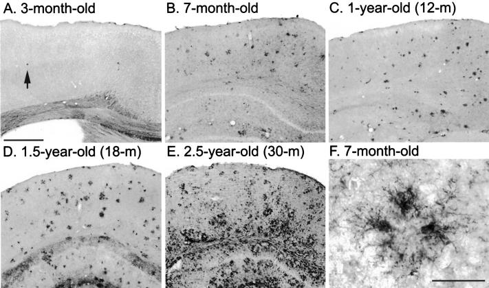
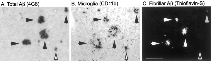
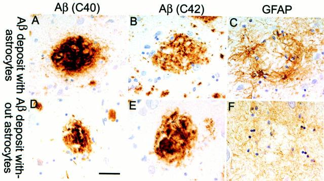
Mutations in the amyloid precursor protein (APP) and presenilin-1 and -2 genes (PS-1, -2) cause Alzheimer's disease (AD). Mice carrying both mutant genes (PS/APP) develop AD-like deposits composed of beta-amyloid (Abeta) at an early age. In this study, we have examined how Abeta deposition is associated with immune responses. Both fibrillar and nonfibrillar Abeta (diffuse) deposits were visible in the frontal cortex by 3 months, and the amyloid load increased dramatically with age. The number of fibrillar Abeta deposits increased up to the oldest age studied (2.5 years old), whereas there were less marked changes in the number of diffuse deposits in mice over 1 year old. Activated microglia and astrocytes increased synchronously with amyloid burden and were, in general, closely associated with deposits. Cyclooxygenase-2, an inflammatory response molecule involved in the prostaglandin pathway, was up-regulated in astrocytes associated with some fibrillar deposits. Complement component 1q, an immune response component, strongly colocalized with fibrillar Abeta, but was also up-regulated in some plaque-associated microglia. These results show: i) an increasing proportion of amyloid is composed of fibrillar Abeta in the aging PS/APP mouse brain; ii) microglia and astrocytes are activated by both fibrillar and diffuse Abeta; and iii) cyclooxygenase-2 and complement component 1q levels increase in response to the formation of fibrillar Abeta in PS/APP mice.


Figures

Figure 1.
Figure 1.
In a longitudinal aging study of PS/APP transgenic mice, Aβ load and distribution were compared in the cerebral cortex, at the level of the amygdala (except A) using antibody 4G8. Aβ deposition in mice younger than 3 months of age was initiated in the frontal cortex (A), but at this age, Aβ deposits were not visible elsewhere (B). By 7 months of age, Aβ deposition in the cerebral cortex and the hippocampus had increased extensively (C and D). After approximately 1 year of age, the total amyloid load remained more consistent (E and F). Scale bar, 1 mm.
Figure 2.
Figure 2.
Fibrillar Aβ as detected by thioflavin-S staining in aging PS/APP transgenic mice was compared in sections taken from the level of the amygdala (except A). Fibrillar Aβ was identified in the frontal cortex of the youngest mice studied, and it increased with total Aβ in aged animals (D–F). Scale bar, 1 mm.
Figure 3.
Figure 3.
Quantitative analysis of amyloid burden in the cortex. The percentage of the frontal cortex area covered by 4G8 immunoreactivity (total Aβ, both fibrillar and diffuse Aβ) or thioflavin-S (fibrillar Aβ only) (open and closed columns, respectively) was measured. Data are presented as mean ± SE (n = 3).
Figure 4.
Figure 4.
The activation of microglia in response to amyloid accumulation was compared in the frontal cortex of PS/APP mouse brains. A few CD11b-immunoreactive activated microglia were observed at 3 months of age (A), and the numbers increased with increasing amyloid burden and age (B–E). Activated microglia were observed surrounding Aβ deposits at all ages (F), although more generalized gliosis was also seen in older mice (E). Scale bars, 500 μm (in A for A–E) and 40 μm (F).
Figure 5.
Figure 5.
Association of activated microglia with fibrillar and nonfibrillar Aβ deposits in 7-month-old PS/APP mouse brain. Two serial sections were stained with anti-Aβ antibody (4G8) (A) and CD11b antibodies (B). CD11b-immunostained section were then counterstained with thioflavin-S to detect fibrillar Aβ (C). CD11b-immunoreactive activated microglia colocalize with both fibrillar Aβ (closed arrows) and thioflavin-S negative (but 4G8 positive) Aβ deposits (open arrow). Scale bar, 100 μm.
Figure 6.
Figure 6.
The activation of plaque-associated astrocytes in the cerebral cortex of PS/APP mice was compared on sections taken at the level of the amygdala (except A). A few activated astrocytes were observed in the vicinity of plaques in the frontal cortex of 3-month-old mice (A), but they were absent from regions devoid of visible Aβ deposits (B). Up to approximately 1 year of age, GFAP-immunoreactive astrocytes were strongly associated with plaques (C and D). In older mice, Aβ-related astrocytosis was extensive except in certain regions of the cortex where the cell bodies and processes appeared more dystrophic or weakly stained (E and F). G and H: GFAP-immunoreactive astrocytes around plaques in the cortex of a 7-month-old mouse (G) and a 2.5-year-old mouse (H). E, F, and H have some background staining on Aβ deposits. Markers (+ and *) indicate the same region in C and G and in F and H, respectively. Scale bars, 200 μm (in A for A–F) and 50 μm (in G for G and H).
Figure 7.
Figure 7.
Association of GFAP astrocytes with plaques in the temporal cortex of a human patient with late-stage AD. The series on the top (A–C) shows an Aβ plaque immunolabeled with antibodies against Aβ40 and Aβ42, respectively (A and B) surrounded by a cluster of reactive astrocytes (C). Bottom panels (D–F) show an Aβ plaque (D and E) which lacks reactive astrocyte cell bodies; however, there is a general meshwork of weakly stained astrocytic processes (F). All panels in this figure were taken from the same section. The sections were counterstained with hematoxylin so that the nuclei stained purple. Scale bar, 50 μm.
Figure 8.
Figure 8.
Induction of COX-2 and its association with Aβ in 7-month-old PS/APP brain (A). Double labeling with COX-2 (red) and GFAP (green) demonstrate colocalization (yellow; A, inset) in astrocytes around plaques. Neuronal COX-2 expression is observed in PS/APP brain (B) as well as in nontransgenic mouse brain (data not shown).
Figure 9.
Figure 9.
Colocalization of C1q with fibrillar Aβ plaques and associated microglia in 12- (A and D) or 7-month-old (C and F) PS/APP mouse brain. C1q-immunostained sections were counterstained with thioflavin-S to detect fibrillar Aβ. To examine the cellular location of C1q, one section was incubated sequentially with fluorescently labeled C1q (C) and F4/80 (F) which was then detected with DAB. C1q appears to colocalize both with plaques and with microglia. Scale bars, 50 μm (in A for A, B, D, and E) and 25 μm (in C for C and F).

References

    1. Hardy J, Duff K, Hardy KG, Perez-Tur J, Hutton M: Genetic dissection of Alzheimer’s disease and related dementias: amyloid and its relationship to tau. Nat Neurosci 1998, 1:355-358 - PubMed
    1. Neve RL, Robakis NK: Alzheimer’s disease: a re-examination of the amyloid hypothesis. Trends Neurosci 1998, 21:15-19 - PubMed
    1. Goedert M: Tau protein and the neurofibrillary pathology of Alzheimer’s disease. Trends Neurosci 1993, 16:460-465 - PubMed
    1. Hsiao K, Chapman P, Nilsen S, Eckman C, Harigaya Y, Younkin S, Yang F, Cole G: Correlative memory deficits, Aβ elevation, and amyloid plaques in transgenic mice. Science 1996, 274:99-102 - PubMed
    1. Duff K, Eckman C, Zehr C, Yu X, Prada CM, Perez-Tur J, Hutton M, Buee L, Harigaya Y, Yager D, Morgan D, Gordon MN, Holcomb L, Refolo L, Zenk B, Hardy J, Younkin S: Increased amyloid-β42(43) in brains of mice expressing mutant presenilin 1. Nature 1996, 383:710-713 - PubMed

Publication types

MeSH terms