Comparative Study
. 2001 Apr;158(4):1379-90.
doi: 10.1016/S0002-9440(10)64089-8.

Replicative senescence of biliary epithelial cells precedes bile duct loss in chronic liver allograft rejection: increased expression of p21(WAF1/Cip1) as a disease marker and the influence of immunosuppressive drugs

Affiliations
Comparative Study

Replicative senescence of biliary epithelial cells precedes bile duct loss in chronic liver allograft rejection: increased expression of p21(WAF1/Cip1) as a disease marker and the influence of immunosuppressive drugs

J G Lunz 3rd et al. Am J Pathol. 2001 Apr.

Abstract

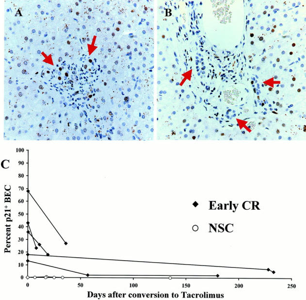
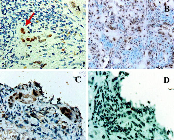
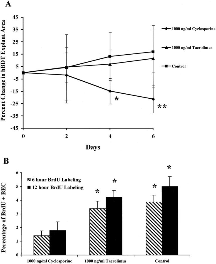
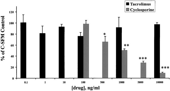
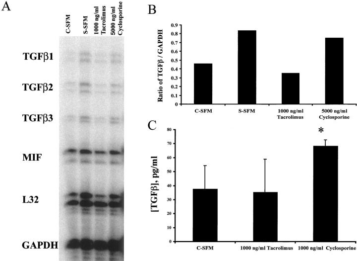
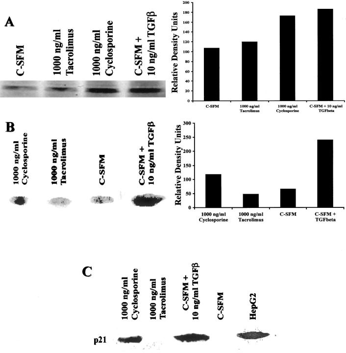
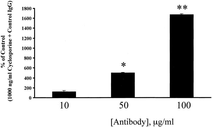
Early chronic liver allograft rejection (CR) is characterized by distinctive cytological changes in biliary epithelial cells (BECs) that resemble cellular senescence, in vitro, and precede bile duct loss. If patients suffering from early CR are treated aggressively, the clinical and histopathological manifestations of CR can be completely reversed and bile duct loss can be prevented. We first tested whether the senescence-related p21(WAF1/Cip1) protein is increased in BECs during early CR, and whether treatment reversed the expression. The percentage of p21+ BECs and the number of p21+ BECs per portal tract is significantly increased in early CR (26 +/- 17% and 3.6 +/- 3.1) compared to BECs in normal liver allograft biopsies or those with nonspecific changes (1 +/- 1% and 0.1 +/- 0.3; P: < 0.0001 and P: < 0.02), chronic hepatitis C (2 +/- 3% and 0.7 +/- 1; P: < 0.0001 and P: < 0.04) or obstructive cholangiopathy (7 +/- 7% and 0.7 +/- 0.6; P: < 0.006 and P: = 0.04). Successful treatment of early CR is associated with a decrease in the percentage of p21+ BECs and the number of p21+ BECs per portal tract. In vitro, nuclear p21(WAF1/Cip1) expression is increased in large and multinucleated BECs, and is induced by transforming growth factor (TGF)-beta. TGF-beta1 also increases expression of TGF-beta receptor II, causes phosphorylation of SMAD-2 and nuclear translocation of p21(WAF1/Cip1), which inhibits BEC growth. Because conversion from cyclosporine to tacrolimus is an effective treatment for early CR, we next tested whether these two immunosuppressive drugs directly influenced BEC growth in vitro. The results show that cyclosporine, but not tacrolimus, stimulates BEC TGF-beta1 production, which in turn, causes BEC mito-inhibition and up-regulation of nuclear p21(WAF1/Cip1). In conclusion, expression of the senescence-related p21(WAF1/Cip1) protein is increased in BECs during early CR and decreases with successful recovery. Replicative senescence accounts for the characteristic BEC cytological alterations used for the diagnosis of early CR and lack of a proliferative response to injury. The ability of cyclosporine to inhibit the growth of damaged BECs likely accounts for the relative duct sparing properties of tacrolimus.


Figures

Figure 1.
Figure 1.
A: High-power photomicrograph of a representative portal tract from a liver allograft recipient with early CR stained for p21WAF1/Cip1 (brown-red nuclei). This biopsy was obtained 8 days before conversion to tacrolimus immunosuppression, when 36% of the BEC nuclei expressed p21WAF1/Cip1 protein (arrows). Note also the enlarged BEC cytoplasm and uneven nuclear spacing, features typical of early CR. B: Repeat biopsy of the same patient, 36 days after conversion to tacrolimus, showed that coincident with clinical improvement, the percentage of p21WAF1/Cip1+ BECs had decreased to 18% and the cytological changes had reversed, as shown in this representative portal tract. C: This graph shows the effect of conversion from cyclosporine to tacrolimus on BEC p21WAF1/Cip1 expression in liver allograft biopsies with either early CR or nonspecific changes. The values at time 0 are the percentages of BECs that stain positive for p21WAF1/Cip1 in the last biopsy obtained while the patient was on cyclosporine. The lines connect consecutive biopsies from the same patient. At time 0, note the increased percentage of p21WAF1/Cip1+ BECs in recipients with early CR compared to those with nonspecific changes. Note also, that the percentage of p21 WAF1/Cip1+ BEC decreases after conversion to tacrolimus in patients with early CR. In contrast, there was no change in the percentage of p21WAF1/Cip1+ BECs in biopsies with nonspecific changes, or in chronic HCV (data not shown).
Figure 2.
Figure 2.
A: Immunohistochemical stain for p21WAF1/Cip1 (brown nuclei) in a liver allograft biopsy with early CR. Note the BEC cytological alterations, including multinucleation (arrow), uneven nuclear spacing, and cellular enlargement, along with the increased p21WAF1/Cip1 nuclear labeling. B: Immunohistochemical stain for p21WAF1/Cip1 in primary cultures of mBEC (brown nuclei). Note the similarity between the damaged BEC in the bile duct with early CR (A and C) and the enlarged senescent mBECs in the primary cultures. Both cell populations show multinucleation and cytoplasmic enlargement and occasional cells show nuclear pyknosis. Note also that both the enlarged cells in the primary BEC cultures and the BECs in damaged bile duct express nuclear p21WAF1/Cip1, but smaller cells do not. C and D: Staining of serial sections from a biopsy with early CR for p21WAF1/Cip1 (C) and Ki-67(Mib-1) (D) shows that the BEC up-regulation of p21WAF1/Cip1 is not associated with mitotic activity.
Figure 3.
Figure 3.
A: The effect of cyclosporine and tacrolimus on the growth of human BECs was tested on human bile-duct tree explants (hBDT), as described in the Materials and Methods. On average, five explant cultures were included in each treatment group. Measurements of culture size were made every 2 days for 6 days. Note the increase in culture size in both the control and tacrolimus-containing media, compared to the significant decrease in size of the cyclosporine-treated cultures (*, P < 0.02; **, P < 0.006 compared to control or tacrolimus-treated cultures). B: To show that the diminished size of the BEC cultures was related to mito-inhibition, the cultures were also labeled with BrdU for either 6 or 12 hours before fixation for anti-BrdU staining. Note the significantly lower percentage of BrdU+ BEC nuclei in the cyclosporine-treated cultures, compared to the others (*, P < 0.01 versus BEC cultures treated with 1,000 ng/ml cyclosporine).
Figure 4.
Figure 4.
Treatment of mBECs kept in C-SFM with increasing concentrations of cyclosporine (100 to 10,000 ng/ml), caused a concentration-dependent decrease in mBECs [3H]-thymidine incorporation after 48 hours, compared to mBECs treated with tacrolimus (0.1 to 10,000 ng/ml). The results shown are representative of at least three separate experiments (*, P < 0.01; **, P < 0.001; ***, P < 0.0003).
Figure 5.
Figure 5.
A: Treatment of mBECs kept in C-SFM with cyclosporine (5,000 ng/ml), but not tacrolimus (1,000 ng/ml) caused an increase of TGF-β mRNA production, as determined by a standard ribonuclease protection assay. If the mBECs were growth arrested by switching to simple serum-free medium (S-SFM) (see Materials and Methods) there was also an increase in TGF-β production. B: Densitometry tracing and comparison to GAPDH mRNA showed that TGF-β mRNA production is higher in the cyclosporine-treated than in the tacrolimus-treated cultures. The determinations were performed 24 hours after the addition of the drugs to the mBEC cultures kept in C-SFM. The results shown are representative of three separate experiments. C: Treatment of mBECs kept in C-SFM with cyclosporine (1,000 ng/ml), but not tacrolimus (1,000 ng/ml) for 48 hours caused an increase of TGF-β protein secretion into the culture supernatant, as determined by a standard enzyme-linked immunosorbent assay (*, P < 0.005).
Figure 6.
Figure 6.
A: Treatment of mBECs kept in C-SFM with cyclosporine (1,000 ng/ml) or TGF-β (10 ng/ml) for 48 hours, but not tacrolimus (1,000 ng/ml) results in up-regulation of the TGF-β receptor type II protein compared to C-SFM controls (left) as determined by Western blotting. Densitometry tracing was used to illustrate the quantitative differences (right). B: TGF-β signaling pathway is mediated by phosphorylation of the transcription factor SMAD-2. In this Western blot (left), total cellular protein was probed for phospho-SMAD-2, which was detected in the mBECs treated with cyclosporine (1,000 ng/ml), but not with tacrolimus (1,000 ng/ml). The positive control consisted of mBECs treated with 10 ng/ml of exogenous TGF-β. Densitometry tracing was used to illustrate the quantitative differences (right). C: In this Western blot, BEC nuclear protein was probed for the cyclin-dependent kinase inhibitor protein, p21WAF1/Cip1, which is elevated 48 hours after treatment with either TGF-β (10 ng/ml; positive control) or cyclosporine (1,000 ng/ml), but not in the control C-SFM or after treatment with tacrolimus (1,000 ng/ml).
Figure 7.
Figure 7.
The simultaneous addition of 1,000 ng/ml cyclosporine and neutralizing anti-TGF-β antibodies to mBECs in C-SFM caused a concentration-dependent increase in [3H]-thymidine incorporation after 48 hours, compared to mBECs treated with 1,000 ng/ml cyclosporine and control nonimmune immunoglobulin. This result further confirms that inhibition of BEC growth, in vitro, is mediated via TGF-β. The results shown are representative of two separate experiments (*, P < 0.002; **, P < 0.0003).

References

    1. Demetris AJ, Fung JJ, Todo S, McCauley J, Jain A, Takaya S, Alessiani M, Abu-Elmagd K, Van Thiel DH, Starzl TE: FK 506 used as rescue therapy for human liver allograft recipients. Transplant Proc 1991, 23:3005-3006 - PMC - PubMed
    1. Freese DK, Snover DC, Sharp HL, Gross CR, Savick SK, Payne WD: Chronic rejection after liver transplantation: a study of clinical, histopathological and immunological features. Hepatology 1991, 13:882-891 - PubMed
    1. Hubscher SG, Buckels JA, Elias E, McMaster P, Neuberger J: Vanishing bile-duct syndrome following liver transplantation—is it reversible? Transplantation 1991, 51:1004-1010 - PubMed
    1. Lautenschlager I, Hockerstedt K, Jalanko H, Loginov R, Salmela K, Taskinen E, Ahonen J: Persistent cytomegalovirus in liver allografts with chronic rejection. Hepatology 1997, 25:190-194 - PubMed
    1. Blakolmer K, Seaberg EC, Batts K, Ferrell L, Markin R, Wiesner R, Detre K, Demetris A: Analysis of the reversibility of chronic liver allograft rejection implications for a staging schema. Am J Surg Pathol 1999, 23:1328-1339 - PubMed

Publication types

MeSH terms