. 2001 Apr;158(4):1451-63.
doi: 10.1016/S0002-9440(10)64096-5.

c-MET expression in myofibroblasts: role in autocrine activation and prognostic significance in lung adenocarcinoma

Affiliations

c-MET expression in myofibroblasts: role in autocrine activation and prognostic significance in lung adenocarcinoma

M Tokunou et al. Am J Pathol. 2001 Apr.

Abstract

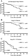
Hepatocyte growth factor (HGF) plays important roles in tumor development and progression. It is currently thought that the main action of HGF is of a paracrine nature: HGF produced by mesenchymal cells acts on epithelial cells that express its receptor c-MET. In this investigation, we explored the significance of c-MET expression in myofibroblasts, both in culture and in patients with lung adenocarcinoma. We first showed that human myofibroblasts derived from primary lung cancer expressed c-MET mRNA and protein by reverse transcription-polymerase chain reaction and Western blot analysis. Proliferation of myofibroblasts was stimulated in a dose-dependent manner by exogenously added recombinant human HGF whereas it was inhibited in a dose-dependent manner by neutralizing antibody to HGF. The addition of HGF in the culture medium stimulated tyrosine phosphorylation of c-MET. The c-MET protein was immunohistochemically detected in myofibroblasts in the invasive area of lung adenocarcinoma. Finally, the prognostic significance of c-MET expression in stromal myofibroblasts was explored in patients with small-sized lung adenocarcinomas. c-MET-positive myofibroblasts were observed in 69 of 131 cases (53%). A significant relationship between myofibroblast c-MET expression and shortened patient survival was observed in a whole cohort of patients including all pathological stages (two-sided P: = 0.0089 by log-rank test) and in patients with stage IA disease (two-sided P: = 0.0019 by log-rank test). These data suggest that the HGF/c-MET system constitutes an autocrine activation loop in cancer-stromal myofibroblasts. This autocrine system may play a role in invasion and metastasis of lung adenocarcinoma.


Figures
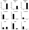
Figure 1.
Figure 1.
Laser-beam microdissection and real-time quantitative RT-PCR analysis. Expression levels of HGF, c-MET, and E-cadherin mRNAs were analyzed by real-time quantitative RT-PCR using total RNA extracted from lung cancer and stromal cells separated by the laser-beam microdissection method. The results were corrected for GAPDH. The laser-beam microdissection and the real-time quantitative RT-PCR analysis were performed as described in Materials and Methods. HGF mRNA expression was found exclusively in the stromal compartment in all of the three cases. E-cadherin mRNA was found only in the cancer cell compartment in tumors 1 and 3. In tumor 2, there was a very low level of E-cadherin mRNA in the stromal compartment, suggesting a minor contamination of epithelial cells. c-MET mRNA was found both in cancer and stromal cell compartments in tumors 1 and 2. Although the c-MET signal in the stromal fraction of tumor 2 was weak, it could not be accounted for by the very low level of epithelial cell contamination.
Figure 2.
Figure 2.
A: RT-PCR analysis of HGF, c-MET, E-cadherin, CD31, and GAPDH mRNA expression in four cultured myofibroblasts derived from primary lung cancer and nontumor lung tissue, and two myofibroblast cell lines. RT-PCR analysis was performed as described in Materials and Methods. Representative results are shown. Lane 1, molecular weight marker; lane 2, N421; lane 3, N515; lane 4, T5062; lane 5, T5162; lane 6, WI38 (myofibroblast cell line); lane 7, MRC5 (myofibroblast cell line); lane 8, MKN28 (positive control for c-MET and E-cadherin); lane 9, HUVEC.SV (positive control for CD31); lane 10, RNA-free sample (negative control for RT-PCR). B: Western blot analysis of c-MET protein in cultured myofibroblasts. Lane 1, MKN45 (positive control for c-MET); lane 2, MRC5 (myofibroblast cell line); lane 3, WI38 (myofibroblast cell line); lane 4, T5162; lane 5, T501; lane 6, T5062; lane 7, N421; lane 8, N425; lane 9, N5162.
Figure 3.
Figure 3.
Scattergram showing expression levels of HGF and c-MET mRNAs in cultured myofibroblasts. HGF and c-MET mRNA levels were determined by real-time quantitative RT-PCR as stated in Material and Methods. The results were corrected for GAPDH and expressed in arbitrary units. Myofibroblasts from lung cancer (T5162, T5062, T421, T425) and from nontumor lung tissue (N421, N425, N515, N5162, N5161) were analyzed.
Figure 4.
Figure 4.
Secretion of HGF in the culture media of myofibroblasts. Cell were incubated for 24 hours in DMEM with 1% FCS and 2 μg of heparin. The concentrations of HGF in the conditioned media were determined by ELISA. The results were corrected for the protein content of the cell layer and expressed as pg/μg protein. The data represent duplicate measurements for each conditioned media.
Figure 5.
Figure 5.
Tyrosine phosphorylation of c-MET in myofibroblasts. Tyrosine phosphorylation of c-MET was examined as stated in Material and Methods by immunoprecipitation with anti-c-MET antibody followed by immunoblotting with anti-phosphotyrosine antibody. Treatment with HGF (25 ng/ml) rapidly induced tyrosine phosphorylation of c-MET. In T425, incubation with neutralizing anti-HGF antibody for 24 hours reduced basal levels of c-MET tyrosine phosphorylation. Lane 1, control; lane 2, 15 minutes after addition of HGF; lane 3, incubation with mouse immunoglobulin (negative control for lane 4); lane 4, 24 hours of incubation with neutralizing antibody against HGF.
Figure 6.
Figure 6.
The effect of HGF/c-MET and other growth factor signaling on proliferation of cultured myofibroblasts. Cell proliferation was measured by the BrdU incorporation assay. A: Proliferation of N515, T5162, and MRC5 myofibroblasts was stimulated by exogenous recombinant HGF. B: Proliferation of N421 myofibroblasts was stimulated by HGF and other growth factors (TGF-β, EGF, aFGF, bFGF, FGF4, PDGF-AB, PDGF-BB) and cytokine (IL-1β). Values represent means ± SD of triplicate measurements. *, P < 0.05 versus control; results were evaluated by one-way analysis of variance followed by Scheffé’s test.
Figure 7.
Figure 7.
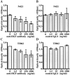
Inhibition of myofibroblast growth by neutralizing antibody against HGF. Cell growth was inhibited by addition of anti-HGF antibody in a dose-dependent manner in N421 and T5062 myofibroblasts (A and C), but not by addition of control IgG1 (B and D). Values represent means ± SD of triplicate measurements. *, P < 0.05 versus control; results were evaluated by one-way analysis of variance followed by Scheffé’s test.
Figure 8.
Figure 8.
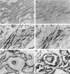
Immunohistochemical analysis of c-MET protein in cancer-stromal myofibroblasts. The c-MET protein expression in cancer-stromal myofibroblasts was evaluated by immunohistochemistry in 131 small-sized lung adenocarcinomas (maximum tumor dimension 2.0 cm or less). A: α-Smooth muscle actin was strongly expressed by almost all cancer stromal cells in the invasive area of a lung adenocarcinoma. C is a higher magnification of A. B: Some of the myofibroblasts that were positive for α-smooth muscle actin were also positive for c-MET. D is a higher magnification of B. Lung adenocarcinoma consisting of c-MET-negative tumor cells is shown here to clearly demonstrate stromal c-MET expression. E: α-Smooth muscle actin was strongly expressed by almost all cancer stromal cells in noninvasive lung adenocarcinoma. F: Only isolated c-MET-positive cells were occasionally observed in the thickened alveolar septa. Most of these cells represented pericytes, endothelial cells, and alveolar myofibroblasts. Original magnifications: ×12.5 (A and B) and ×100 (C–F).
Figure 9.
Figure 9.
Kaplan-Meier survival curves according to the presence or absence of c-MET expression in cancer stromal myofibroblasts for all pathological stage patients with small-sized lung adenocarcinoma (A) as well as for patients with stage IA (B) or stage IB∼IV (C) disease. The survival differences between the two curves were analyzed using the log-rank test.

References

    1. Nakamura T: Structure and function of hepatocyte growth factor. Prog Growth Factor Res 1991, 3:67-85 - PubMed
    1. Gak E, Taylor WG, Chan AM, Rubin JS: Processing of hepatocyte growth factor to the heterodimeric form is required for biological activity. FEBS Lett 1992, 311:17-21 - PubMed
    1. Mizuno K, Tanoue Y, Okano I, Harano T, Takada K, Nakamura T: Purification and characterization of hepatocyte growth factor (HGF)-converting enzyme: activation of pro-HGF. Biochem Biophys Res Commun 1994, 198:1161-1169 - PubMed
    1. Jiang W, Hiscox S, Matsumoto K, Nakamura T: Hepatocyte growth factor/scatter factor, its molecular, cellular and clinical implications in cancer. Crit Rev Oncol Hematol 1999, 29:209-248 - PubMed
    1. Nakamura T, Nishizawa T, Hagiya M, Seki T, Shimonishi M, Sugimura A, Tashiro K, Shimizu S: Molecular cloning and expression of human hepatocyte growth factor. Nature 1989, 342:440-443 - PubMed

Publication types

MeSH terms