. 2001 Apr;158(4):1473-80.
doi: 10.1016/s0002-9440(10)64098-9.

Expression of leukemia-associated antigen, JL1, in bone marrow and thymus

Affiliations

Expression of leukemia-associated antigen, JL1, in bone marrow and thymus

Y K Shin et al. Am J Pathol. 2001 Apr.

Abstract

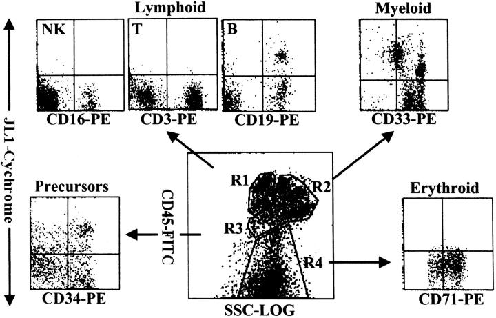
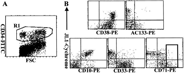
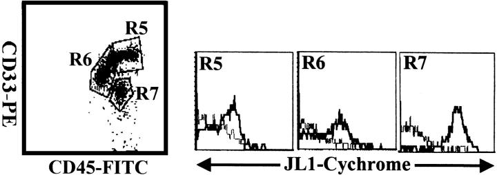
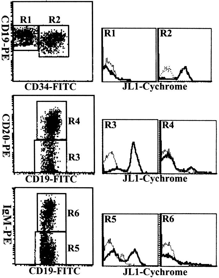
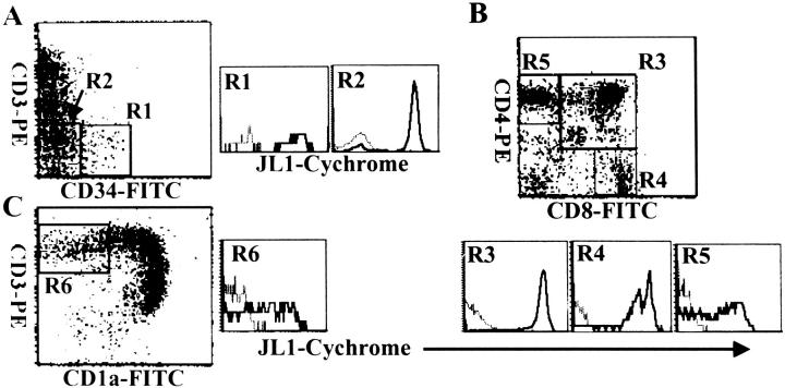
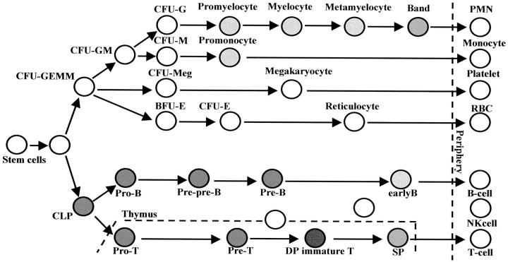
The identification of immunophenotypic markers with restricted expression has long been a critical issue in diagnostic and therapeutic advances for acute leukemias. We previously developed a monoclonal antibody against a new thymocyte surface antigen, JL1, and showed that JL1 is expressed in the majority of acute leukemia cases. In this study, using multiparameter flow cytometric analyses, we found that JL1 was uniquely expressed in subpopulations of normal bone marrow (BM) cells, implying the association of JL1 with the differentiation and maturation process. Although CD34(+) CD10(+) lymphoid precursors and some of maturing myeloid cells express JL1, neither CD34(+) CD38(-/lo) nor CD34(+) AC133(+) noncommitted pluripotent stem cells do. As for the myeloid precursors, CD34(+) CD33(+) cells do not express JL1. During lymphopoiesis, JL1 on the earliest lymphoid precursors disappear in the CD20(+) sIgM(+) stage of B-cell development or after CD1a down-regulation in thymocytes. Despite the highly restricted expression of JL1 in normal BM cells, most of the leukemias express JL1 irrespective of their immunophenotypes. These results indicate that JL1 is not only a novel differentiation antigen of hematopoietic cells, but also a leukemia-associated antigen. Therefore, we suggest that JL1 be a candidate molecule in acute leukemia for the diagnosis and immunotherapy that spares the normal BM stem cells.


Figures

Figure 1.
Figure 1.
Flow cytometric analysis of JL1 expression of BM leukocytes. MNCs obtained by density-gradient separation were labeled with biotinylated JL1 mAb and SA-cychrome, followed by staining with CD45-FITC in combination with CD3-PE, CD19-PE, CD16-PE, CD33-PE, CD71-PE, or CD34-PE, respectively. Normal BM cells were divided into four normal populations by CD45-side scatter analysis and the expressions of JL1 as well as lineage-specific markers were measured on the corresponding regions. R1, lymphocytes and lymphoblasts; R2, myelomonocytic lineage; R3, early precursors; R4, erythroid cells including nucleated red blood cells.
Figure 2.
Figure 2.
The expression of JL1 molecules on CD34+ BM progenitor cells. BM cells were labeled with biotinylated JL1 mAb and SA-cychrome, followed by staining with CD34-FITC in combination with CD38-PE, AC133-PE, CD10-PE, CD33-PE, or CD71-PE, respectively. CD34+ cells were gated (R1; A) and JL1 expression in R1 gate was compared on each of the progenitor cells (B). A square indicates CD71bright cell population.
Figure 3.
Figure 3.
Three-color flow cytometric analysis of JL1 expression in myeloid population of G-CSF-treated BM MNCs. MNCs obtained by density-gradient separation were labeled with biotinylated JL1 mAb and SA-cychrome, followed by staining with CD45-FITC and CD33-PE. Each lineage cell cluster was painted on the CD45 versus side-scatter plot and JL1 expression was compared. Dashed lines represent isotype control levels for each population. R5, promonocytes and monocytes; R6, promyelocytes, myelocytes, and metamyelocytes; R7, band forms.
Figure 4.
Figure 4.
Three-color cytometric analysis of the JL1 expression of B-lineage cells in BM MNCs. BM cells were labeled with biotinylated JL1 mAb and SA-cychrome, followed by staining with CD34-FITC and CD19-PE, or CD19-FITC in the combination with CD20-PE and IgM-PE, respectively. Dashed lines represent isotype control levels for each of the colored populations.
Figure 5.
Figure 5.
Three-color flow cytometric analysis of the JL1 expression in human thymocytes. Thymocytes were labeled with biotinylated JL1 mAb and SA-cychrome, followed by staining with CD34-FITC and CD3-PE (A), or CD8-FITC and CD4-PE (B), or CD1a-FITC and CD3-PE (C). Dashed lines represent isotype control levels for each of the colored populations.
Figure 6.
Figure 6.
Expression pattern of JL1 antigen during hematopoiesis. Filled circle represents JL1-expressing populations in BM or thymus, and the degree of its darkness reflects the levels of expression of JL1 molecule.

References

    1. Compana D, Pui CH: Detection of minimal residual disease in acute leukemia: methodologic advances and clinical significance. Blood 1995, 85:1416-1434 - PubMed
    1. Caron PC, Scheinberg DA: Immunotherapy for acute leukemias. Curr Opin Oncol 1994, 6:14-22 - PubMed
    1. Jurcic JG, Caron PC, Scheinberg DA: Monoclonal antibody therapy of leukemia and lymphoma. Adv Pharmacol 1995, 33:287-314 - PubMed
    1. Maloney DG: Advances in immunotherapy of hematologic malignancies. Curr Opin Hematol 1998, 5:237-243 - PubMed
    1. Maloney DG: Advances in the immunotherapy of hematologic malignancies: cellular and humoral approaches. Curr Opin Hematol 1999, 6:222-228 - PubMed

Publication types