. 2001 Jun 15;276(24):21303-10.
doi: 10.1074/jbc.M102316200. Epub 2001 Apr 4.

Identification of common binding sites for calmodulin and inositol 1,4,5-trisphosphate receptors on the carboxyl termini of trp channels

Affiliations

Identification of common binding sites for calmodulin and inositol 1,4,5-trisphosphate receptors on the carboxyl termini of trp channels

J Tang et al. J Biol Chem. .

Abstract

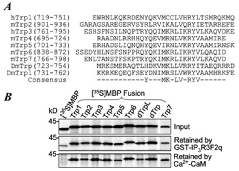
Homologues of Drosophila Trp (transient receptor potential) form plasma membrane channels that mediate Ca(2+) entry following the activation of phospholipase C by cell surface receptors. Among the seven Trp homologous found in mammals, Trp3 has been shown to interact with and respond to IP(3) receptors (IP(3)Rs) for activation. Here we show that Trp4 and other Trp proteins also interact with IP(3)Rs. The IP(3)R-binding domain also interacts with calmodulin (CaM) in a Ca(2+)-dependent manner with affinities ranging from 10 nm for Trp2 to 290 nm for Trp6. In addition, other binding sites for CaM and IP(3)Rs are present in the alpha but not the beta isoform of Trp4. In the presence of Ca(2+), the Trp-IP(3)R interaction is inhibited by CaM. However, a synthetic peptide representing a Trp-binding domain of IP(3)Rs inhibited the binding of CaM to Trp3, -6, and -7 more effectively than that to Trp1, -2, -4, and -5. In inside-out membrane patches, Trp4 is activated strongly by calmidazolium, an antagonist of CaM, and a high (50 microm) but not a low (5 microm) concentration of the Trp-binding peptide of the IP(3)R. Our data support the view that both CaM and IP(3)Rs play important roles in controlling the gating of Trp-based channels. However, the sensitivity and responses to CaM and IP(3)Rs differ for each Trp.


Figures

Fig. 1
Fig. 1. Localization of IP3R-binding domain and CaM-binding sites at the C terminus of mTrp4
A, diagram of mTrp4α and its fragments included in the EBFP or MBP fusion proteins. Numbers in parentheses indicate the positions of the fragments. Thick and thin lines denote that the binding to CaM is positive and negative, respectively. Shaded boxes in the full-length mTrp4α indicate the locations of trans-membrane (TM) segments. B, representative binding results showing the autoradiograms of input 35S-labeled EBFP-Trp4 C-terminal (CT), MBP-Trp4 N-terminal (NT) fusion proteins, EBFP (E), and MBP (M) and those retained by GST, GST-IP3R3F2q, and CaM. A picture of a Coomassie Blue-stained gel displayed below the second panel shows the amount of GST and GST fusion protein used. C and D, additional binding results showing that two C-terminal regions of mTrp4α (CT1 and CT2) (C) and Trp4Cc (D) bind to both IP3R3F2q and Ca2+/CaM.
Fig. 2
Fig. 2. Colocalization of binding sites for Ca2+/CaM and IP3R3F2q on Trp4CT1
A, compositions of EBFP or MBP fusion proteins containing subfragments of Trp4Cc. Positions in the full-length mTrp4α are shown in parentheses. Relative intensities of the fusion proteins retained by Ca2+/CaM and GST-IP3R3F2q are indicated by the plus and minus signs and are summarized from 2–4 binding experiments. B and C show binding results from representative experiments.
Fig. 3
Fig. 3. Determination of binding sites for Ca2+/CaM and IP3R3F2q on Trp4CT2α
A, diagrams of mTrp4α and mTrp4β and compositions of MBP fusion proteins containing subfragments of Trp4CT2. Positions in respect to the full-length mTrp4α are shown in parentheses. CT2β contains the CT2 fragment of mTrp4β and lacks the 84-amino acid region (shown as a dashed line in MBP-CT2β and black box in mTrp4α). Thick and thin lines denote that the binding to CaM is positive and negative, respectively. B–D show binding results from representative experiments. Like Trp4CT2p, Trp4CT2h–o did not bind to GST-IP3R3F2q (not shown).
Fig. 4
Fig. 4. The common CaM/IP3R binding site is present in all Trp proteins
A, amino acid compositions of mammalian Trp1–7, Drosophila Trp (DmTrp), and TrpL (DmTrpL) present in the MBP fusion proteins. Positions for these sequences in the full-length proteins are shown in parentheses. 35S-Labeled MBP fusion proteins were tested for binding to GST-IP3R3F2q and to Ca2+/CaM as described under “Materials and Methods.” Representative binding results are shown in B.
Fig. 5
Fig. 5. All IP3Rs interact with the C terminus of Trp3
A, amino acid compositions of IP3R1, IP3R2, and IP3R3 included in the GST fusion proteins. B, amino acid compositions of subregions of IP3R3F2q included in the GST fusion proteins. Positions in the full-length proteins are indicated in parentheses. 35S-Labeled hTrp3 C terminus (T3CT, Asn725–Glu848) was tested for binding to the GST-IP3R fusion proteins as described under “Materials and Methods.” Representative binding results are shown in C. Upper panels show [35S]T3CT retained by the GST fusion proteins as revealed by autoradiography, while lower panels show the amount of GST fusion proteins used as revealed by staining with Coomassie Blue.
Fig. 6
Fig. 6. Competition between CaM and IP3R3 for binding to the Trp CIRB domains
A, CaM inhibits Trp4 binding to IP3R3. Varying concentrations of CaM were included in the binding reactions for 35S-labeled MBP-T4Cw and GST-IP3R3F2q. The binding buffer contained 70 μM free Ca2+. After washing, bound [35S]MBP-T4Cw was separated by SDS-polyacrylamide gel electrophoresis. The left panel shows an autoradiogram from a representative experiment, while the right panel shows averages of results of phosphorimaging analysis of the relative amount of [35S]T4Cw retained from three experiments. The curve is the least-square fit of the equation, y = 1/(1 + [P]/IC50), where y is relative binding, [P] is CaM concentration, and IC50 = 2.1 μM is the concentration that causes 50% inhibition. B, representative experiments show that 20 μM CaM inhibits the binding of IP3R3F2q to the CIRB domain of Trp1, −2, −5, −6, and −7 in a Ca2+-dependent manner. 10 mM HEDTA and EGTA were used to buffer [Ca2+] to 50 μM and 0 (<10 nM), respectively. C, binding of Trp4CT2c to IP3R3F2q was inhibited by 20 μM CaM. [Ca2+] = 50 μM. D, peptide F2v inhibits binding of CaM to the CIRB domain of Trp1–7. Varying concentrations of peptide F2v were included in the binding reaction for 35S-labeled CaM and GST fusion proteins containing the CIRB sequence of Trp1–7 or the second CaM-binding site of Trp4 (CT2k, 781–814). Positions of Trp sequences included in the GST fusion proteins are indicated in parentheses. The binding buffer contained 70 μM free Ca2+. After washing, bound [35S]CaM was separated by SDS-polyacrylamide gel electrophoresis and revealed by autoradiography. IC50 values were determined from results of phosphorimaging analysis of the relative amount of [35S]CaM retained by each Trp fragment from two or three experiments. The percentages of inhibitions for different concentrations of F2v were fitted with the equation as in A, except that [P] is the concentration of the peptide.
Fig. 7
Fig. 7. Peptide F2v and CMZ stimulate channel activity in inside-out patches excised from Trp4
Inside-out patches were excised from untransfected HEK293 cells (control) or two stable cell lines expressing mTrp4α (T4–60 and T4–1) to a bath solution that contained either no Ca2+ (A) or 18 μM Ca2+ (B) as described under “Materials and Methods.” Peptide F2v at 5 or 50 μM (A) and CMZ at 1 or 10 μM (B) were applied to the cytoplasmic side of the membrane by perfusion. Bar graphs show averages ± S.E. of the mean current (sampled from periods of 400 s) at −40 mV from the numbers of patches indicated in parentheses. Representative traces for control and T4–60 cells at basal level and 50 μM F2v (A) or 10 μM CMZ (B) are shown on the right. Dashed lines indicate closed level. *, p < 0.01; **, p < 0.05 different from basal level by Student’s t test.

Similar articles

Cited by

References

    1. Putney JW., Jr Cell Calcium. 1986;7:1–12. - PubMed
    1. Putney JW., Jr Cell Calcium. 1990;11:611–624. - PubMed
    1. Berridge MJ. Biochem J. 1995;312:1–11. - PMC - PubMed
    1. Clapham DE. Cell. 1995;80:259–268. - PubMed
    1. Birnbaumer L, Zhu X, Jiang M, Boulay G, Peyton M, Vannier B, Brown D, Platano D, Sadeghi H, Stefani E, Birnbaumer M. Proc Natl Acad Sci U S A. 1996;93:15195–15202. - PMC - PubMed

Publication types

MeSH terms

LinkOut - more resources