. 2001 Apr 15;166(8):5279-85.
doi: 10.4049/jimmunol.166.8.5279.

Anergy induction by dimeric TCR ligands

Affiliations

Anergy induction by dimeric TCR ligands

H Appel et al. J Immunol. .

Abstract

T cells that recognize particular self Ags are thought to be important in the pathogenesis of autoimmune diseases. In multiple sclerosis, susceptibility is associated with HLA-DR2, which can present myelin-derived peptides to CD4(+) T cells. To generate molecules that target such T cells based on the specificity of their TCR, we expressed a soluble dimeric DR2-IgG fusion protein with a bound peptide from myelin basic protein (MBP). Soluble, dimeric DR2/MBP peptide complexes activated MBP-specific T cells in the absence of signals from costimulatory or adhesion molecules. This initial signaling through the TCR rendered the T cells unresponsive (anergic) to subsequent activation by peptide-pulsed APCs. Fluorescent labeling demonstrated that anergic T cells were initially viable, but became susceptible to late apoptosis due to insufficient production of cytokines. Dimerization of the TCR with bivalent MHC class II/peptide complexes therefore allows the induction of anergy in human CD4(+) T cells with a defined MHC/peptide specificity.


Figures

FIGURE 1
FIGURE 1
Kinetics of initial T cell proliferation induced by DR2/ MBP-IgG. Activation of clone Ob.2F3 by soluble DR2/MBP-IgG (20 μg/ ml) was compared with stimulation with rIL-2 or peptide-pulsed MNC from a DR2+ donor. Mouse IgG2a was used as a negative control. Soluble DR2/MBP-IgG induced initial T cell activation with similar kinetics as peptide-pulsed MNC. DR2/MBP-IgG or mouse IgG2a (negative control) was added at 20 μg/ml to 105 T cells/well. For stimulation with MBP (85–99) peptide, MNC from a DR2+ donor were pulsed overnight with MBP (85–99) (1 μM), washed, and cocultured with T cells (105 MNC and 105 T cells/well). rIL-2 was used at a concentration of 5 U/ml. T cell proliferation was quantitated by [3H]thymidine incorporation. The x-axis reflects the total time of T cells in culture, including the thymidine pulse.
FIGURE 2
FIGURE 2
Anergy induction by DR2/MBP-IgG. T cells specific for the DR2/MBP peptide complex become anergic following pretreatment with soluble or immobilized DR2/MBP-IgG. A, T cells from clone Ob.2F3 were treated with either rIL-2 or soluble DR2/MBP-IgG (20 μg/ml in medium without rIL-2) for 96 h. T cells were then washed three times and resus-pended in medium without rIL-2. T cells (105) were then assayed by co-culture with 5 × 10⁴ peptide-pulsed B cells, and T cell proliferation was measured after 48 h by [3H]thymidine incorporation. B, T cells from clone Ob.2F3 were treated with immobilized DR2/MBP-IgG or anti-CD3 for 96 h. Molecules were immobilized in a 96-well plate by overnight incubation of 200 ng/well in 50 μl of 100 mM bicarbonate, pH 9.6. Wells were washed twice with PBS, and 105 T cells were added to each well. After 96 h, T cells were tested for the ability to proliferate to peptide-pulsed B cells, as described above. The same experiments were performed with T cell clone Ob.1A12, using soluble (C) and immobilized molecules (D).
FIGURE 3
FIGURE 3
A, Dose dependence of anergy induction. T cells from clone Ob.2F3 were treated with soluble DR2/MBP-IgG (1.25–20 μg/ml in medium without rIL-2) or with rIL-2 for 96 h. T cells were then washed three times and resuspended in medium without rIL-2. T cells (105) were assayed by coculture with 5 × 10⁴ peptide-pulsed B cells, and T cell proliferation was measured after 48 h by [3H]thymidine incorporation. B, Anergy induction by DR2/MBP-IgG in the presence of MNCs. T cells (5 × 10⁴/well) were pretreated with 20 μg/ml of soluble DR2/MBP-IgG in the absence or presence of irradiated MNC from a DR2+ donor (5 × 10⁴ cells/well). After 96 h, T cells were washed twice in RPMI, resuspended in medium without rIL-2, and cocultured with peptide-pulsed B cells (105 T cells, 5 × 10⁴ B cells/well). After 48 h of culture, T cell proliferation was measured by [3H]thymidine incorporation. These experiments demonstrate that DR2/MBP-IgG can also induce anergy when the molecules can be captured by Fc receptors expressed on APC.
FIGURE 4
FIGURE 4
Anergic T cells proliferate in response to rIL-2. Anergic T cells are viable and proliferate in response to rIL-2, but not to stimulation via the TCR. T cells (clone Ob.2F3) were pretreated with 20 μg/ml of soluble DR2/MBP-IgG for 96 h in medium without rIL-2. T cells were washed and cocultured with peptide-pulsed B cells in the presence or absence of rIL-2 (5 U/ml). T cell proliferation was measured by [3H]thymidine incorporation after 48 h of culture.
FIGURE 5
FIGURE 5
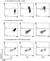
Survival and proliferation of anergic T cells in the presence of rIL-2. T cells from clone Ob.2F3 were pretreated with 20 μg/ml of soluble DR2/MBP-IgG for 96 h and labeled with 0.5 μM CFSE for 1 h at 37°C. T cells were washed three times with PBS, resuspended in medium with rIL-2, and plated on 96-well plates in the presence of peptide-pulsed DR2+ B cells. After 24, 60, or 108 h, T cells were stained with Alexa 594-labeled annexin V and analyzed by FACS. In the presence of IL-2, the anergic T cells were viable at all three time points, indicated by the fact that they remained annexin V negative. Proliferation in response to the rIL-2 was most apparent at 108 h, as indicated by a stepwise loss of CFSE labeling.
FIGURE 6
FIGURE 6
Anergic T cells become susceptible to late apoptosis following stimulation with B cells and peptide. T cells (clone Ob.2F3) were pretreated with 20 μg/ml of soluble DR2/MBP-IgG (A), peptide-pulsed B cells (B), or 20 μg/ml of soluble DR2/MBP-IgG plus 10 μg/ml of soluble anti-CD28 (C). After 96 h, T cells were labeled with CSFE and cocultured with peptide-pulsed B cells. Following 24, 60, and 108 h, T cells were stained with Alexa 594-labeled annexin V, followed by FACS analysis. The gate was set such that cellular debris was excluded from analysis. A, After 24 h of stimulation with peptide-pulsed B cells, the majority of anergic T cells were viable (91.7% annexin V negative). However, after 60 and 108 h, large numbers of anergic T cells were apoptotic. The annexin V-positive population comprised 38% and 53.1% of gated T cells at 60 and 108 h, respectively. B, In contrast, T cells that had been pretreated with peptide-pulsed B cells proliferated vigorously following re-stimulation. Proliferation was already apparent at 24 h by a loss of CFSE-staining intensity. Significant cell division was also evident at 60 h, and T cells remained viable throughout the experiment. C, T cells pretreated with soluble DR2/MBP-IgG in the presence of soluble anti-CD28 (clone 3D10) were not anergic and showed extensive proliferation and little apoptosis.

References

    1. Zamvil SS, Steinman L. The T lymphocyte in experimental allergic encephalomyelitis. Annu Rev Immunol. 1990;8:579. - PubMed
    1. Steinman L. Multiple sclerosis: a coordinated immunological attack against myelin in the central nervous system. Cell. 1996;85:299. - PubMed
    1. Goverman J, Woods A, Larson L, Weiner LP, Hood L, Zaller DM. Transgenic mice that express a myelin basic protein-specific T cell receptor develop spontaneous autoimmunity. Cell. 1993;72:551. - PubMed
    1. McDevitt HO. The role of MHC class II molecules in susceptibility and resistance to autoimmunity. Curr Opin Immunol. 1998;10:677. - PubMed
    1. Nepom GT, Erlich H. MHC class-II molecules and autoimmunity. Annu Rev Immunol. 1991;9:493. - PubMed

Publication types

MeSH terms