

Identification of attenuated Yersinia pseudotuberculosis strains and characterization of an orogastric infection in BALB/c mice on day 5 postinfection by signature-tagged mutagenesis
- PMID: 11292689
- PMCID: PMC98225
- DOI: 10.1128/IAI.67.5.2779-2787.2001
Identification of attenuated Yersinia pseudotuberculosis strains and characterization of an orogastric infection in BALB/c mice on day 5 postinfection by signature-tagged mutagenesis
Abstract
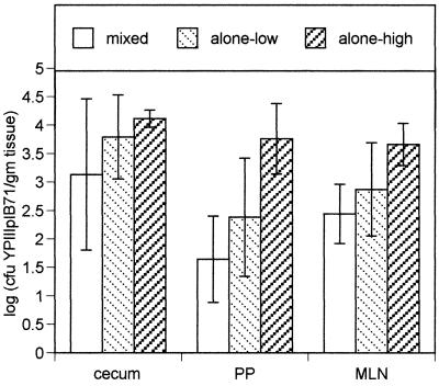
Yersinia pseudotuberculosis localizes to the distal ileum, cecum, and proximal colon of the gastrointestinal tract after oral infection. Using signature-tagged mutagenesis, we isolated 13 Y. pseudotuberculosis mutants that failed to survive in the cecum of mice after orogastric inoculation. Twelve of these mutants were also attenuated for replication in the spleen after intraperitoneal infection, whereas one strain, mutated the gene encoding invasin, replicated as well as wild-type bacteria in the spleen. Several mutations were in operons encoding components of the type III secretion system, including components involved in translocating Yop proteins into host cells. This indicates that one or more Yops may be necessary for survival in the gastrointestinal tract. Three mutants were defective in O-antigen biosynthesis; these mutants were also unable to invade epithelial cells as efficiently as wild-type Y. pseudotuberculosis. Several other mutations were in genes that had not previously been associated with growth in a host, including cls, ksgA, and sufl. In addition, using Y. pseudotuberculosis strains marked with signature tags, we counted the number of different bacterial clones that were present in the cecum, mesenteric lymph nodes, and spleen 5 days postinfection. We find barriers in the host animal that limit the number of bacteria that succeed in reaching and/or replicating in the mesenteric lymph nodes and spleen after breaching the gut mucosa.
Figures
References
-
- Allaoui A, Schulte R, Cornelis G R. Mutational analysis of the Yersinia enterocolitica virC operon: characterization of yscE, F, G, I, J, K required for Yop secretion and yscH encoding YopR. Mol Microbiol. 1995;18:343–355. - PubMed
-
- Blanchin-Roland S, Blanquet S, Schmitter J M, Fayat G. The gene for Escherichia coli diadenosine tetraphosphatase is located immediately clockwise to folA and forms an operon with ksgA. Mol Gen Genet. 1986;205:515–522. - PubMed
Publication types
MeSH terms
Substances
LinkOut - more resources
Full Text Sources
Other Literature Sources
Molecular Biology Databases
