. 2001 May;69(5):3057-66.
doi: 10.1128/IAI.69.5.3057-3066.2001.

Antigenic variation of Anaplasma marginale: major surface protein 2 diversity during cyclic transmission between ticks and cattle

Affiliations

Antigenic variation of Anaplasma marginale: major surface protein 2 diversity during cyclic transmission between ticks and cattle

A F Barbet et al. Infect Immun. 2001 May.

Abstract

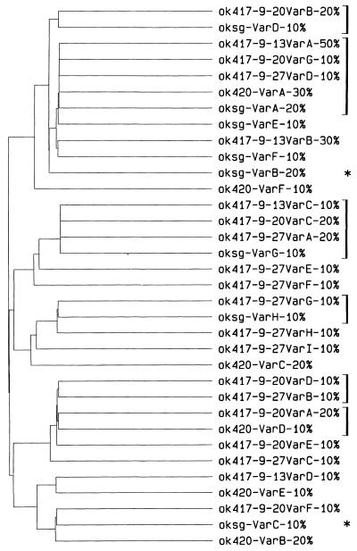
The rickettsial pathogen Anaplasma marginale expresses a variable immunodominant outer membrane protein, major surface protein 2 (MSP2), involved in antigenic variation and long-term persistence of the organism in carrier animals. MSP2 contains a central hypervariable region of about 100 amino acids that encodes immunogenic B-cell epitopes that induce variant-specific antibodies during infection. Previously, we have shown that MSP2 is encoded on a polycistronic mRNA transcript in erythrocyte stages of A. marginale and defined the structure of the genomic expression site for this transcript. In this study, we show that the same expression site is utilized in stages of A. marginale infecting tick salivary glands. We also analyzed the variability of this genomic expression site in Oklahoma strain A. marginale transmitted from in vitro cultures to cattle and between cattle and ticks. The structure of the expression site and flanking regions was conserved except for sequence that encoded the MSP2 hypervariable region. At least three different MSP2 variants were encoded in each A. marginale population. The major sequence variants did not change on passage of A. marginale between culture, acute erythrocyte stage infections, and tick salivary glands but did change during persistent infections of cattle. The variant types found in tick salivary glands most closely resembled those present in bovine blood at the time of acquisition of infection, whether infection was acquired from an acute or from a persistent rickettsemia. These variations in structure of an expression site for a major, immunoprotective outer membrane protein have important implications for vaccine development and for obtaining an improved understanding of the mechanisms of persistence of ehrlichial infections in humans, domestic animals, and reservoir hosts.


Figures

FIG. 1
FIG. 1
Derivation of cyclically transmitted A. marginale populations for analysis of sequence diversity in the polycistronic msp2 expression site.
FIG. 2
FIG. 2
PLOTSIMILARITY profiles of nucleotide sequence variability in msp2 expression sites show greatest variability in the central region of the msp2 gene. (a) The 7 different populations from the Oklahoma strain of A. marginale described in Fig. 1 are compared; (b) Florida, Idaho, and Oklahoma strain acute bloodstream populations are compared. A similarity score of 1.0 indicates identical sequence in a sliding window of 10 nucleotides; a decreasing score from 1.0 to 0.0 indicates increasing variation.
FIG. 3
FIG. 3
A polycistronic RNA transcript containing the msp2 gene is present in A. marginale-infected salivary glands from D. variabilis and D. andersoni ticks. RT-PCR analysis of isolated RNA from infected salivary glands with AB198 as the RT primer, AB765 and AB766 as primary PCR primers, and AB192 and AB783 as secondary (nested) PCR primers. A 2.1-kbp product was specifically amplified in reactions containing reverse transcriptase enzyme (+) but was not present in control reactions without reverse transcriptase (−). This 2.1-kbp band hybridized to an orf2 probe (arrow). Cloning and sequencing of the 2.1-kbp product demonstrated that it contained sequence from msp2, orf2, orf3, and orf4. Low-molecular-weight hybridizing bands are also present in RNA (lanes labeled +), which may represent amplified products from partially degraded A. marginale RNA.
FIG. 4
FIG. 4
Structure of msp2 and orf2 to orf4 in genomic DNA of Florida, South Idaho, and Oklahoma strains of A. marginale. Southern blots of Florida (F), South Idaho (I), Oklahoma acute erythrocyte stage (Oe), or culture stage (Oc) genomic DNA digested with the restriction enzyme FspI and hybridized with probes specific for either msp2, orf2, orf3, or orf4 (probe is shown at bottom of figure). FspI cleaves 41 nucleotides 5′ to orf4 and 268 nucleotides 3′ to msp2 to release a fragment of 3.76 kbp that contains the complete polycistronic msp2 expression site sequence (see Fig. 2) from all genomic DNAs. Molecular size standards (Std) are shown in the left lane of each blot. Multiple msp2-related sequences are present in genomic DNA of all strains; only msp2 sequences located in the expression site are contiguous with orf2, orf3, and orf4.
FIG. 5
FIG. 5
Multiple different msp2 variants are present in the polycistronic expression site in each population of A. marginale. The major variant type is conserved during passage of A. marginale between culture, acute erythrocyte stage infection, and tick salivary glands but is not conserved in persistent cattle infections. The expression site was amplified by PCR using primers which annealed 288 bp 3′ to the termination codon of msp2 (AB752) and to the intercistronic sequence between orf3 and orf4 (AB750) to generate a product of 2.9 kbp from A. marginale genomic DNA that contained msp2, orf2, and orf3. The PCR product was cloned in pCR-XL-TOPO vector (Invitrogen), and independent colonies containing a 2.9-kbp insert were selected for sequencing of cloned plasmid DNA. The hypervariable region of the msp2 gene was sequenced on both strands in seven independent clones derived by PCR amplification from genomic DNA of each of the A. marginale populations described in Fig. 1. DNA sequences were translated to amino acids, and the different variant sequences were aligned with PILEUP. The proportion of each sequence variant in that population is indicated in brackets; e.g., the major sequence variant detected in cultured A. marginale was variant A, which was found in three of seven independent clones of the expression site. Identical amino acids shared between all variants are indicated by a dash and are shown on the bottom row of the alignment. Variant types present in different A. marginale populations are indicated by identical symbols to the left of the sequence alignments.
FIG. 6
FIG. 6
Msp2 expression site variability increases in persistent cattle infection. The variability of each population was calculated over the hypervariable region of the msp2 expression site using the seven independent clones sampled from each of the seven A. marginale populations in Fig. 1, the sequence alignment in Fig. 5, and the following formula: number of different amino acids at a given position/frequency of the most common amino acid at that position. For example, at position 7 in population okcul there is H (histidine) in six clones and Y (tyrosine) in one clone of the msp2 expression site, for a variability of 2/0.857 (=2.3). Values were obtained similarly for all variable positions in the alignment and added, to obtain a total population variability for okcul of 163.8.
FIG. 7
FIG. 7
Circulating msp2 expression site variants in persistently infected cattle are those transmitted by ticks to naive cattle. Ten independent clones of the msp2 expression site were sampled from each of five different populations of A. marginale. These populations were derived from persistently infected calf PA417 just prior to tick feeding (ok417-9-13 variants), during tick feeding (ok417-9-20 variants), and just after tick feeding (ok417-9-27 variants) and also from salivary glands of D. variabilis that acquired infection from calf PA417 (oksg variants) and from the acute bloodstream rickettsemia that was transmitted by those ticks to calf PA420 (ok420 variants). The 50 clones were sequenced over the hypervariable region, translated to amino acids, and compared using PILEUP as explained in Fig. 5. The figure is the dendrogram output from PILEUP that shows the clustering of similar and identical sequences in the different A. marginale populations. Brackets to the right identify identical variants in different A. marginale populations; e.g., variant type ok417-9-13VarA was found in 50% (5 of 10) of the expression site clones sampled from calf PA417 prior to tick feeding. Identical variant types were found in other samplings of calf PA417 during and after tick feeding, in salivary glands of ticks that acquired infection from calf PA417, and in the acute rickettsemia that was transmitted to calf PA420. The asterisk indicates that three of ten expression site clones from tick salivary gland stages of A. marginale had changes that would lead to synthesis of truncated MSP2. In two of ten (oksg-VarB) clones the change was the same base substitution that introduced a termination codon into the hypervariable region; in oksg-VarC it was a single base deletion that changed the reading frame leading to termination shortly after the deletion. We cannot exclude the possibility that these changes are due to PCR and/or cloning errors; it is also possible that one result of recombination mechanisms can be variant types with truncated MSP2.

References

    1. Alleman A R, Kamper S M, Viseshakul N, Barbet A F. Analysis of the Anaplasma marginale genome by pulsed-field electrophoresis. J Gen Microbiol. 1993;139:2439–2444. - PubMed
    1. Andrew H R, Norval R A. The carrier status of sheep, cattle and African buffalo recovered from heartwater. Vet Parasitol. 1989;34:261–266. - PubMed
    1. Barbet A F, Kamper S M. The importance of mosaic genes to trypanosome survival. Parasitol Today. 1993;9:63–66. - PubMed
    1. Barbet A F, Lundgren A, Yi J, Rurangirwa F R, Palmer G H. Antigenic variation of Anaplasma marginale by expression of MSP2 mosaics. Infect Immun. 2000;68:6133–6138. - PMC - PubMed
    1. Barbet A F, Palmer G H, Myler P J, McGuire T C. Characterization of an immunoprotective protein complex of Anaplasma marginale by cloning and expression of the gene coding for polypeptide Am105L. Infect Immun. 1987;55:2428–2435. - PMC - PubMed

Publication types

Substances

LinkOut - more resources