. 2001 May;69(5):3067-72.
doi: 10.1128/IAI.69.5.3067-3072.2001.

BrkA protein of Bordetella pertussis inhibits the classical pathway of complement after C1 deposition

Affiliations

BrkA protein of Bordetella pertussis inhibits the classical pathway of complement after C1 deposition

M G Barnes et al. Infect Immun. 2001 May.

Abstract

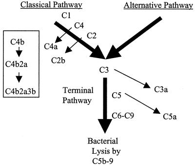
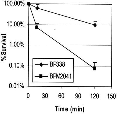
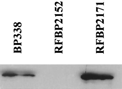
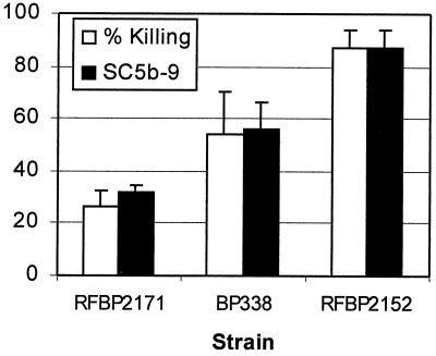
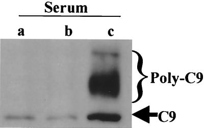
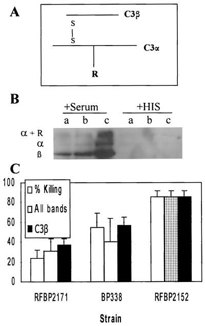
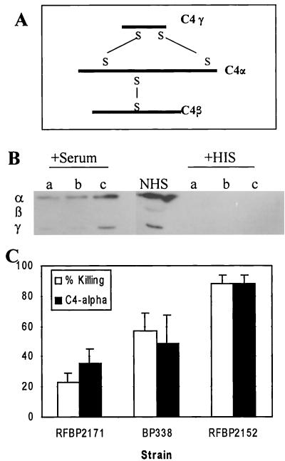
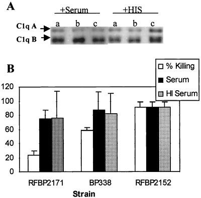
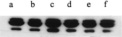
Bordetella pertussis produces a 73-kDa protein, BrkA (Bordetella resistance to killing), which inhibits the bactericidal activity of complement. In this study we characterized the step in the complement cascade where BrkA acts, using three strains: a wild-type strain, a strain containing an insertional disruption of brkA, and a strain containing two copies of the brkA locus. Following incubation with 10% human serum, killing was greatest for the BrkA mutant, followed by that for the wild-type strain, while the strain with two copies of brkA was the most resistant. Complement activation was monitored by enzyme-linked immunosorbent assay (ELISA) or Western blotting. ELISAs for SC5b-9, the soluble membrane attack complex, showed that production of SC5b-9 was greatest with the brkA mutant, less with the wild type, and least with the strain containing two copies of brkA. Deposition of complement proteins on the bacteria was monitored by Western blotting. A decrease in deposition on the bacteria of C4, C3, and C9 corresponded with decreased complement sensitivity. Deposition of C1, however, was not affected by the presence of BrkA. These studies show that BrkA inhibits the classical pathway of complement activation and prevents accumulation of deposited C4.


Figures

FIG. 1
FIG. 1
The complement cascade. Both the classical and alternative pathways of complement activation are represented, although details are given only for the classical pathway. The nonactivated complement proteins are indicated above and to the right of the wide arrows, and the arrows pointing from them show the product that is released during activation. The smaller flowchart in the box indicates the membrane-bound products and complexes formed by the classical pathway on the bacterial surface.
FIG. 2
FIG. 2
Survival of B. pertussis in complement. BP338 and BPM2041 were incubated with 10% human serum for 15 (P < 0.0038) or 120 (P < 0.0012) min. Percent survival is calculated relative to a non-killing heat-inactivated serum control. The results are the average and standard error of four separate trials. BPM2041 is the BrkA mutant; BP338 is the wild type.
FIG. 3
FIG. 3
BrkA expression. A representative Western blot showing the variation in expression of BrkA protein from the three strains used for the killing assays and deposition assays. BP338, wild type; RFBP2152, BrkA mutant. RFBP2171 contains two copies of brkA.
FIG. 4
FIG. 4
Production of the soluble membrane attack complex, SC5b-9, relative to bacterial killing. Empty bars represent percent killing; solid bars represent SC5b-9 production as determined by ELISA relative to RFBP2152 with standard deviation. Three separate experiments were performed. Values for RFBP2152 are significantly different from those for BP338 (P = 0.05) and RFBP2171 (P < 0.0005).
FIG. 5
FIG. 5
C9 deposition. Bacteria were incubated in 10% serum for 15 min. Equal numbers of cells were subjected to Western blotting with a goat anti-human C9 antibody. Strains were RFBP2171 (a), BP338 (b), and RFBP2152 (c). The bracket indicates the region of poly-C9 deposition; the arrow indicates mono-C9 deposition.
FIG. 6
FIG. 6
C3 deposition. (A) C3 is composed of an α chain and β chain connected by disulfide bonds (S—S). The α chain can bind covalently to the bacterial surface (represented by R). (B) Bacteria were incubated in 10% normal human serum or heat-inactivated serum (HIS) for 15 min and were analyzed by Western blotting under reducing conditions using goat anti-human C3 antibodies. RFBP2171 (a), BP338 (b), and RFBP2152 (c) were used. The α and β chains were determined by a comparison to bacterium-free serum run as a size control. α + R indicates larger bands than those seen in a serum control. (C) Densitometry was performed on the Western blots, and results are graphed in arbitrary units relative to RFBP2152 with standard error from four separate experiments. “All bands” indicates densitometry of all bands observed in the Western blot, while “C3β” indicates densitometry of only the lower, C3β band. Densitometry of “All bands” and “C3β ” for RFBP2152 was significantly higher than for BP338 (P < 0.04 and 0.03, respectively) or for RFBP2171 (P < 0.0005 and 0.004, respectively).
FIG. 7
FIG. 7
C4 deposition. (A) C4 is composed of three chains, α, β and γ, connected by disulfide bonds (S—S). (B) Bacteria were incubated in 10% serum or heat-inactivated serum (HIS) for 15 min and were analyzed by Western blotting under reducing conditions with goat anti-human C4 antibodies. RFBP2171 (a), BP338 (b), and RFBP2152 (c) were tested. NHS, normal human sera. The three chains were determined by a comparison with bacterium-free serum that was run as a size control. (C) Densitometry was performed, and results are graphed as arbitrary units relative to RFBP2152 with standard error of three separate experiments. Densitometry results for RFBP2152 were significantly higher than for BP338 (P < 0.02) or for RFBP2171 (P < 0.03).
FIG. 8
FIG. 8
C1 deposition. (A) Bacteria were incubated in 10% serum or heat-inactivated serum (HIS) for 15 min and were analyzed under reducing conditions using goat anti-human C1q antibodies. RFBP2171 (a), BP338 (b), and RFBP2152 (c) were used. (B) Densitometry was performed, and results are graphed as arbitrary units relative to RFBP2152 with standard error determined from five independent experiments. Differences were not significant. HI, heat inactivated.
FIG. 9
FIG. 9
Deposition of purified C1. Bacteria were incubated with purified C1 (a and d), purified C1 with purified IgG (b and e), or heat-inactivated purified C1 (c and f) for 15 min and were analyzed under reducing conditions using goat anti-human C1q antibodies. a to c, BP338; d to f, RFBP2171.

References

    1. Akesson P, Sjoholm A G, Bjorck L. Protein SIC, a novel extracellular protein of Streptococcus pyogenes interfering with complement function. J Biol Chem. 1996;271:1081–1088. - PubMed
    1. Berggard K, Johnsson E, Mooi F R, Lindahl G. Bordetella pertussis binds the human complement regulator C4BP: role of filamentous hemagglutinin. Infect Immun. 1997;65:3638–3643. - PMC - PubMed
    1. Bock S C, Skriver K, Nielsen E, Thogersen H C, Wiman B, Donaldson V H, Eddy R L, Marrinan J, Radziejewska E, Huber R, et al. Human C1 inhibitor: primary structure, cDNA cloning, and chromosomal localization. Biochemistry. 1986;25:4292–4301. - PubMed
    1. Brzezinski R M, Boutelet-Bochan H, Person R E, Fantel A G, Juchau M R. Catalytic activity and quantitation of cytochrome P-450 2E1 in prenatal human brain. J Pharmacol Exp Ther. 1999;289:1648–1653. - PubMed
    1. Burns S M, Hull S I. Comparison of loss of serum resistance by defined lipopolysaccharide mutants and an acapsular mutant of uropathogenic Escherichia coli O75:K5. Infect Immun. 1998;66:4244–4253. - PMC - PubMed

Publication types

MeSH terms