. 2001 May;69(5):3175-80.
doi: 10.1128/IAI.69.5.3175-3180.2001.

Extensive Mycobacterium bovis BCG infection of liver parenchymal cells in immunocompromised mice

Affiliations

Extensive Mycobacterium bovis BCG infection of liver parenchymal cells in immunocompromised mice

J W Mills et al. Infect Immun. 2001 May.

Abstract

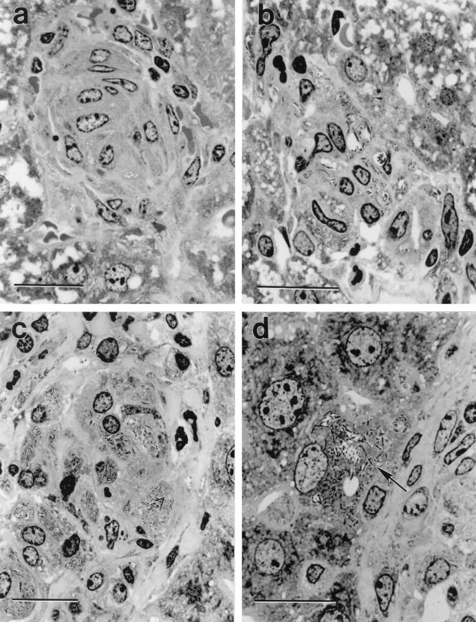
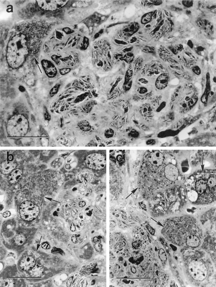
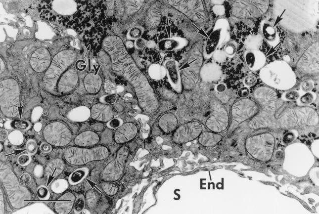
A histologic study was performed on the livers of wild-type (WT), severe combined immunodeficient (SCID), hydrocortisone acetate (HC)-treated WT, and HC-treated SCID mice infected intravenously with 10(5) CFU of Mycobacterium bovis BCG. It was found that infection progressed faster in SCID mice than in WT mice and that HC treatment caused exacerbation of infection in both types of mice. In all cases infection in the liver was confined to granulomas that were populated predominantly by macrophages. Higher levels of infection in HC-treated SCID mice, but not HC-treated WT mice, were associated with extensive infection and destruction of parenchymal cells at the margins of granulomas. The results indicate that in the absence of T-cell-mediated immunity and of HC-sensitive T-cell-independent defense mechanisms, macrophages are incapable of restricting BCG growth and of confining infection to their cytoplasm. Consequently, BCG bacilli are released into the extracellular environment, where they are ingested by neighboring parenchymal cells.


Figures

FIG. 1
FIG. 1
Crystal violet-stained, 2-μm-thick plastic section of a liver granuloma. The granuloma in the WT mouse (a) is compact and comprised predominantly of concentrically arranged macrophages, whereas the granuloma in the HC-treated WT mouse (b) is comprised of a loose collection of macrophages containing large numbers of BCG bacilli. The granuloma in the SCID mouse (c) is also comprised predominantly of macrophages that are heavily infected with BCG. The higher power micrograph of the edge of a granuloma in a SCID mouse (d) shows a hepatocyte at the margin of the granuloma (arrow) heavily infected with BCG. Bars represent 20 μm. Magnifications: ×1,060 (a), ×1,280 (b), ×1020 (c), and ×1,280 (d).
FIG. 2
FIG. 2
Crystal violet-stained, 2-μm-thick plastic sections of liver granulomas in SCID mice treated with HC. The micrograph of a whole granuloma (a) shows very heavily infected macrophages containing dense rafts of BCG and a heavily infected hepatocyte (arrow) at the margin of the granuloma. Note that the infected hepatocyte is the same large size as the uninfected hepatocyte below it and is much lager than macrophages in the granuloma. Each of the other two micrographs (b and c) was selected to show heavily infected hepatocytes (arrows) at the margins of granulomas. Bars represent 20 μm. Magnifications: ×1,360 (a) and ×1,190 (b and c).
FIG. 3
FIG. 3
Electron micrograph of an infected hepatocyte in an HC-treated SCID mouse. The hepatocyte was situated at the edge of a granuloma (out of view at the top) and next to a sinusoid (S) marginated by typical endothelium (E). The hepatocyte is heavily infected with BCG bacilli (arrows) that are enclosed in phagosomes. Mitochondria and glycogen deposits (G) typical of hepatocytes are clearly visible. Bars, 2 μm.

References

    1. Ashwell J D, Lu F W M, Vacchio M S. Glucocorticoids in T cell development and function. Annu Rev Immunol. 2000;18:309–345. - PubMed
    1. Cross C C, Mercer K L. Cell and tissue ultrastructure. A functional perspective. W. H. Oxford, England: Freeman and Company; 1993.
    1. Drouet C, Shakhov A N, Jongeneel C V. Enhancers and transcription factors controlling the inducibility of tumor necrosis factor-alpha promoter in primary macrophages. J Immunol. 1991;147:1694–1670. - PubMed
    1. Ellis R S, Zabrowany L A. Safer staining method for acid fast bacilli. J Clin Pathol. 1993;46:559–560. - PMC - PubMed
    1. Flynn J L, Goldstein M M, Treibold K J, Koller B, Bloom B R. Major histocompatibility complex class I-restricted T cells are required for resistance to Mycobacterium tuberculosis infection. Proc Natl Acad Sci USA. 1992;89:12013–12017. - PMC - PubMed

Publication types

LinkOut - more resources