. 2001 May;69(5):3343-9.
doi: 10.1128/IAI.69.5.3343-3349.2001.

Fas-mediated apoptosis of neutrophils in sera of patients with infection

Affiliations

Fas-mediated apoptosis of neutrophils in sera of patients with infection

I E Nwakoby et al. Infect Immun. 2001 May.

Abstract

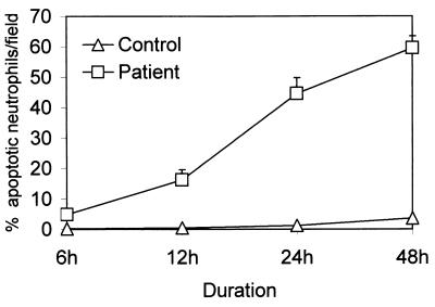
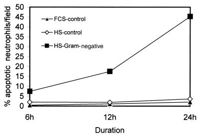
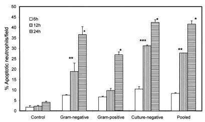
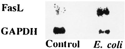
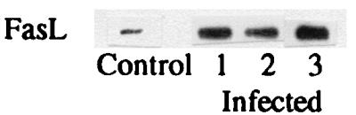
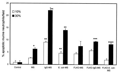
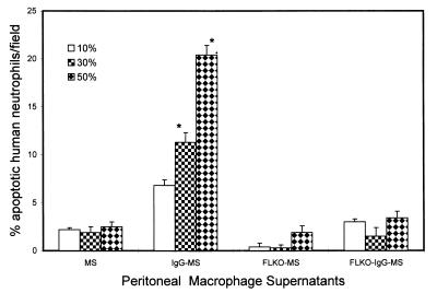
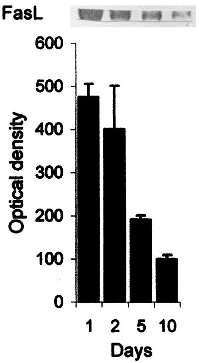
In the presence of infection, neutropenia is considered to be a marker of poor prognosis; conversely, neutrophilia may not be a determinant of a better prognosis. Since apoptotic neutrophils are compromised functionally, we evaluated the effect of infection on neutrophil apoptosis. The rate of apoptosis was greater for neutrophils isolated from patients with infection than for healthy controls. Escherichia coli did not directly modulate the rate of neutrophil apoptosis. However, sera from infected patients promoted (P < 0.001) neutrophil apoptosis. Interestingly, the sera of patients with different types of infection (gram negative, gram positive, or culture negative) exerted a more or less identical response on neutrophil apoptosis. Sera of infected patients showed a fivefold greater content of FasL compared to controls. Moreover, anti-FasL antibody partly attenuated the infected-serum-induced neutrophil apoptosis. In in vitro studies, E. coli enhanced monocyte FasL expression. Moreover, conditioned media prepared from activated macrophages from control mice showed enhanced apoptosis of human as well as mouse neutrophils. On the contrary, conditioned media prepared from activated macrophages isolated from FasL-deficient mice induced only a mild degree of neutrophil apoptosis. These results suggest that neutrophils in patients with infection undergo apoptosis at an accelerated rate. Infection not only promoted monocyte expression of FasL but also increased FasL content of the serum. Because the functional status of apoptotic cells is compromised, a significant number of neutrophils may not be participating in the body's defense. Since neutrophils play the most important role in innate immunity, their compromised status in the presence of infection may transfer the host defense burden from an innate response to acquired immunity. The present study provides some insight into the lack of correlation between neutrophilia and the outcome of infection.


Figures

FIG. 1
FIG. 1
Morphologic evaluation of neutrophil apoptosis. Neutrophils were isolated from healthy subjects and infected patients and were incubated in medium containing 10% pooled human sera for 24 h. At the end of the incubation period, cells were stained with H-33342 and propidium iodide. (A) Neutrophils isolated from healthy subjects; (B) neutrophils isolated from infected subjects. Apoptotic neutrophils show bright fluorescence, whereas necrosed cells are stained pink.
FIG. 2
FIG. 2
Time course effect on aging neutrophils in vitro. Neutrophils were isolated from healthy subjects (open triangles) and infected patients (open squares) and were incubated in medium containing 10% FCS serum for 6, 12, 24, and 48 h. At the end of the incubation period, cells were stained with H-33342 and propidium iodide. Results (means ± SEM) are from six series of experiments, each carried out in triplicate.
FIG. 3
FIG. 3
Effect of sera on neutrophil apoptosis. Neutrophils isolated from healthy subjects were incubated in media containing either 10% FCS (FCS-control), 10% HS (sera from healthy subjects) (HS control) or 10% gram-negative HS (sera from gram-negative-infected patients) for 6, 12, and 24 h. At the end of the incubation period, cells were stained with H-33342 and propidium iodide. Results (means ± SEM) are from six series of experiments, each carried out in triplicate.
FIG. 4
FIG. 4
Effect of gram-negative and gram-positive sera on neutrophil apoptosis. Neutrophils isolated from healthy subjects were incubated in media containing either 10% HS (sera from healthy subjects, control), 10% gram-negative sera (sera from gram-negative infected patients), 10% gram-positive sera (sera from gram-positive infected patients), 10% culture-negative sera (sera from infected culture-negative patients), or 10% pooled sera (infected patients) for 6, 12, and 24 h. At the end of the incubation period, cells were stained with H-33342 and propidium iodide. Results (means ± SEM) are from three series of experiments, each carried out in triplicate. ∗, P < 0.001 compared with the respective control; ∗∗, P < 0.01 compared with the respective control; ∗∗∗, P < 0.001 compared with the respective control.
FIG. 5
FIG. 5
Effect of E. coli on monocyte FasL expression. The upper lane shows monocyte mRNA expression of FasL in control and E. coli-treated conditions. The lower lane shows monocyte mRNA expression of GAPDH under identical conditions.
FIG. 6
FIG. 6
Western blots showing FasL content in serum for control and infected patients. Sera of infected patients showed a fivefold increase of FasL compared with control sera.
FIG. 7
FIG. 7
Effect of activated peritoneal macrophage (derived from either normal mice or FasL-deficient mice) supernatants on murine neutrophil apoptosis. Results (means ± SEM) are from four sets of experiments, each carried out in triplicate. FLKO-IgG-MS induced apoptosis in a lower percentage of murine neutrophils than IgG-MS. Similarly, E. coli-MS triggered apoptosis in a greater percentage of murine neutrophils than FLKO-E. coli-MS. This effect of macrophage supernatants was dose dependent, indicating the presence of an apoptotic factor. ∗, P < 0.01 compared with the respective control; ∗∗, P < 0.001 compared with the respective control; ∗∗∗, P < 0.001 compared with IgG-MS; ∗∗∗∗, P < 0.001 compared with E. coli-MS.
FIG. 8
FIG. 8
The effect of activated peritoneal macrophage (derived from either normal mice or FasL-deficient mice) supernatants (10, 30, and 50%) on human neutrophils. Results (means ± SEM) are from four sets of experiments, each carried out in triplicate. IgG-MS induced apoptosis in a greater percentage of human neutrophils than FLKO-IgG-MS. ∗, P < 0.001 compared with respective MS, FLKO-MS, and FLKO-IgG-MS.
FIG. 9
FIG. 9
Serum FasL kinetics in two patients with gram-negative infection. Sera were collected from two patients on days 1, 2, 5, and 10 and FasL contents were measured. Results (means ± SEM) are from two patients. A representative Western blot showing FasL content in serum at different time periods is shown at the top.

References

    1. Badley A D, Dockrell D, Simpson M, Schut R, Lynch D H, Leibson P, Paya C V. Macrophage-dependent apoptosis of CD4+ T lymphocytes from HIV-infected individuals is mediated by FasL and tumor necrosis factor. J Exp Med. 1997;185:55–64. - PMC - PubMed
    1. Badley A D, McElhinny J A, Leibson P J, Lynch D H, Alderson M R, Paya C V. Upregulation of Fas ligand expression by human immunodeficiency virus in human macrophages mediates apoptosis of uninfected T lymphocytes. J Virol. 1996;70:199–206. - PMC - PubMed
    1. Brown S B, Savill J. Phagocytosis triggers macrophage release of Fas ligand and induces apoptosis of bystander neutrophils. J Immunol. 1999;162:480–485. - PubMed
    1. Colotta F, Polentarutti N, Sozzani S, Montovani A. Modulation of granulocyte survival and programmed cell death by cytokines and bacterial products. Blood. 1992;80:2012–2020. - PubMed
    1. Cox G. Glucocorticoid treatment inhibits apoptosis in human neutrophils. Separation of survival and activation outcomes. J Immunol. 1995;154:4719–4725. - PubMed

Publication types