. 2001 May;69(5):3350-8.
doi: 10.1128/IAI.69.5.3350-3358.2001.

Visualizing pneumococcal infections in the lungs of live mice using bioluminescent Streptococcus pneumoniae transformed with a novel gram-positive lux transposon

Affiliations

Visualizing pneumococcal infections in the lungs of live mice using bioluminescent Streptococcus pneumoniae transformed with a novel gram-positive lux transposon

K P Francis et al. Infect Immun. 2001 May.

Abstract

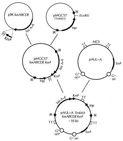
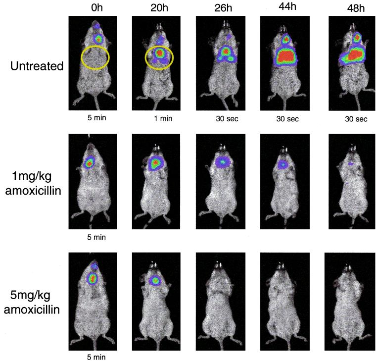
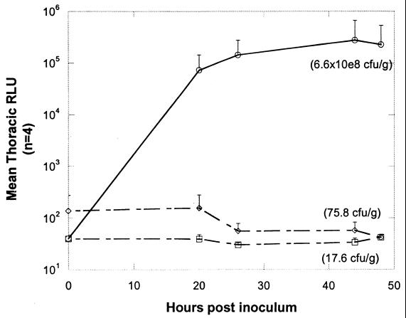
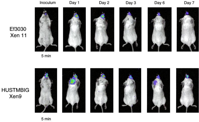
Animal studies with Streptococcus pneumoniae have provided valuable models for drug development. In order to monitor long-term pneumococcal infections noninvasively in living mice, a novel gram-positive lux transposon cassette, Tn4001 luxABCDE Km(r), that allows random integration of lux genes onto the bacterial chromosome was constructed. The cassette was designed so that the luxABCDE and kanamycin resistance genes were linked to form a single promoterless operon. Bioluminescence and kanamycin resistance only occur in a bacterial cell if this operon has transposed downstream of a promoter on the bacterium's chromosome. S. pneumoniae D39 was transformed with plasmid pAUL-A Tn4001 luxABCDE Km(r), and a number of highly bioluminescent colonies were recovered. Genomic DNA from the brightest D39 strain was used to transform a number of clinical S. pneumoniae isolates, and several of these strains were tested in animal models, including a pneumococcal lung infection model. Strong bioluminescent signals were seen in the lungs of the animals containing these pneumococci, allowing the course and antibiotic treatment of the infections to be readily monitored in real time in the living animals. Recovery of the bacteria from the animals showed that the bioluminescent signal corresponded to the number of CFU and that the lux construct was highly stable even after several days in vivo. We believe that this lux transposon will greatly expand the ability to evaluate drug efficacy against gram-positive bacteria in living animals using bioluminescence.


Figures

FIG. 1
FIG. 1
Construction of plasmid pAUL-A Tn4001 luxABCDE Kmr. For details, see Materials and Methods. IR, inverted repeat; EmR, erythromycin resistance gene; tnp, transposase gene; TT, transcription terminator; MCS, multiple cloning site.
FIG. 2
FIG. 2
Effects of kanamycin concentration on selection of promoter strength. S. pneumoniae D39 Xen 7 was grown to mid-exponential phase, and 103 CFU were plated on chocolate agar plates supplemented with various concentrations of kanamycin. Open squares, total CFU on the plates; solid circles, ratio of light to total CFU. The numbers of CFU are the averages for five plates at each kanamycin concentration.
FIG. 3
FIG. 3
RLU emitted by bioluminescent S. pneumoniae strains during in vitro growth. Three hundred microliters of overnight culture was inoculated into 30 ml of BHI and grown at 37°C in 5% CO2. At 1-h intervals, both the OD600 (open circles) and the number of RLU × 106 (solid squares) from a 1-ml culture volume were determined for each of the strains. The experiment was repeated three times for each strain.
FIG. 4
FIG. 4
Bioluminescent S. pneumoniae A66.1 Xen 10 in a mouse pneumococcal lung model, with and without antibiotic treatment. Twelve mice were inoculated with approximately 106 CFU of S. pneumoniae A66.1 Xen 10. The mice were divided into three groups of four animals each, and two of these groups were treated with amoxicillin at 1 or 5 mg/kg given subcutaneously at 0, 18, 24, and 42 h postinfection; the third group of animals were left untreated as controls. Mice were imaged using an IVIS CCD camera (Xenogen Corporation) at the indicated time points postinfection. Total photon emission from the ventral thoracic region of each mouse (areas inside the yellow circle) was quantified using the LivingImage software package (Xenogen Corporation). Imaging time was 5 min unless otherwise shown.
FIG. 5
FIG. 5
Graphic representation of mean thoracic bioluminescence (RLU) from the pneumococcus-infected mice, untreated and treated with amoxicillin, shown in Fig. 4. Each point represents the average bioluminescence from all surviving mice in each treatment group (starting with four mice in each group). ○, untreated animals; ◊, treated (5 mg/kg) animals; □, treated (1 mg/kg) animals. The average CFU per gram of lung tissue for each group of animals at 48 h (time of sacrifice) is given in parentheses.
FIG. 6
FIG. 6
Bioluminescent S. pneumoniae Ef3030 Xen9 and HUSTMBIG Xen 11 in a mouse nasopharygeal model. Mice were infected intranasally with approximately 106 CFU by placing 20 μl of bacterial suspension on the nares and allowing the mice to inhale the inoculum. Mice were imaged using an IVIS CCD camera (Xenogen Corporation) at the indicated time points postinfection. Imaging time was 5 min in each case.

References

    1. Appelbaum P C. Antimicrobial resistance in Streptococcus pneumoniae: an overview. Clin Infect Dis. 1992;15:77–83. - PubMed
    1. Austrian R. Some aspects of the pneumococcal carrier state. J Antimicrob Chemother. 1986;18(Suppl. A):35–45. - PubMed
    1. Briles D E, Paton J C, Swiatlo E, Nahm M H. Pneumococcal vaccines. In: Fischetti V A, et al., editors. Gram-positive pathogens. Washington, D.C.: American Society for Microbiology; 2000. pp. 244–250.
    1. Buckley N D, Lee L N, LeBlanc D J. Use of a novel mobilizable vector to inactivate the scrA gene of Streptococcus sobrinus by allelic replacement. J Bacteriol. 1995;177:5028–5034. - PMC - PubMed
    1. Caparon M. Genetics of group A streptococci. In: Fischetti V A, et al., editors. Gram-positive pathogens. Washington, D.C.: American Society for Microbiology; 2000. pp. 53–65.

LinkOut - more resources