. 2001 Apr;12(4):847-62.
doi: 10.1091/mbc.12.4.847.

RAC1 regulates adherens junctions through endocytosis of E-cadherin

Affiliations
Free PMC article

RAC1 regulates adherens junctions through endocytosis of E-cadherin

N Akhtar et al. Mol Biol Cell. 2001 Apr.
Free PMC article

Abstract

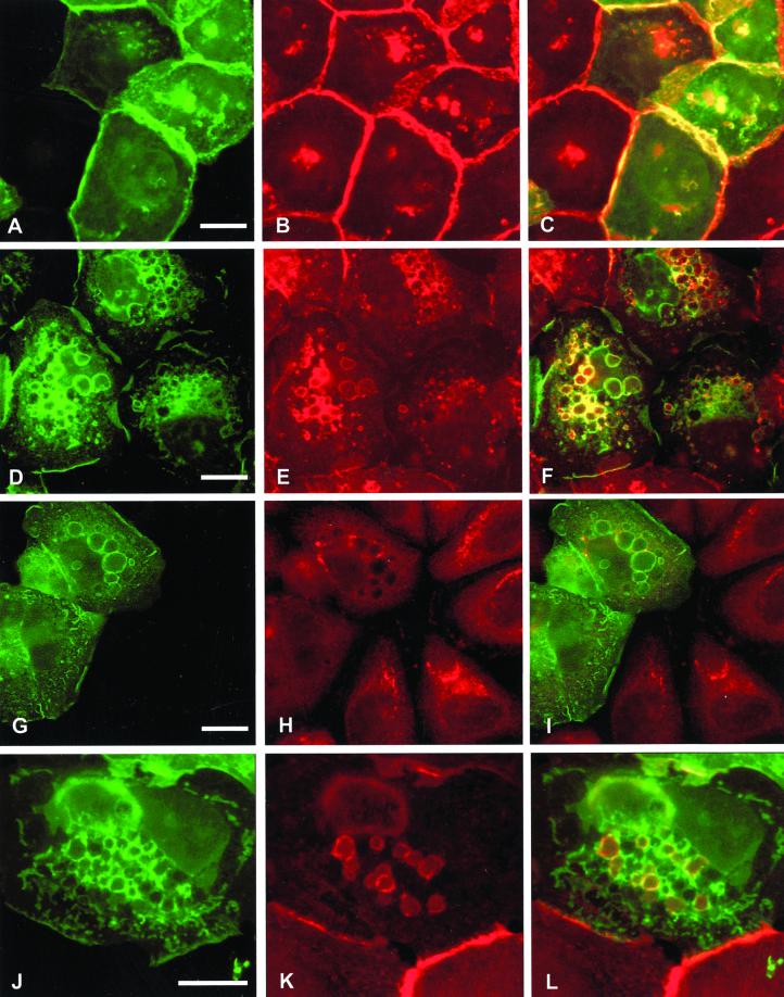
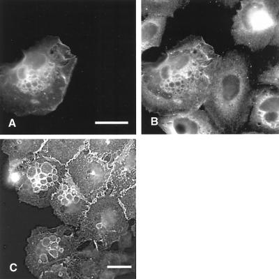
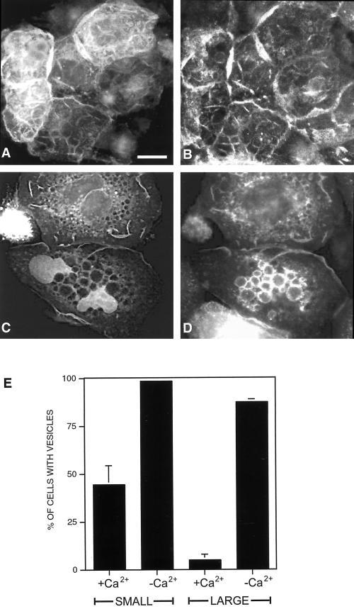
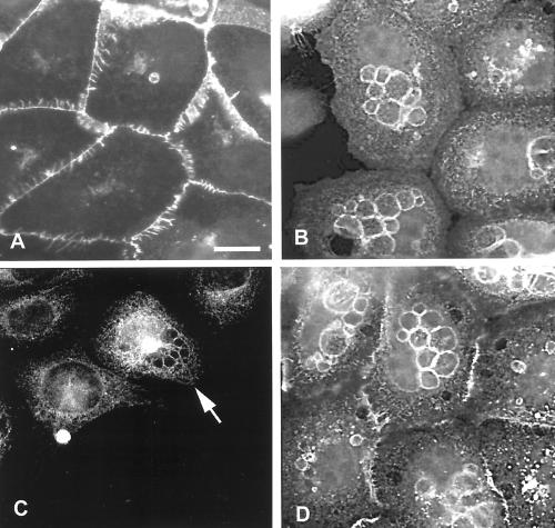
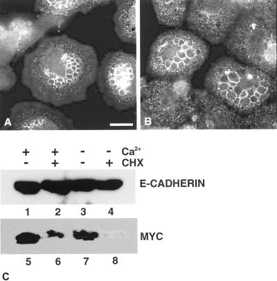
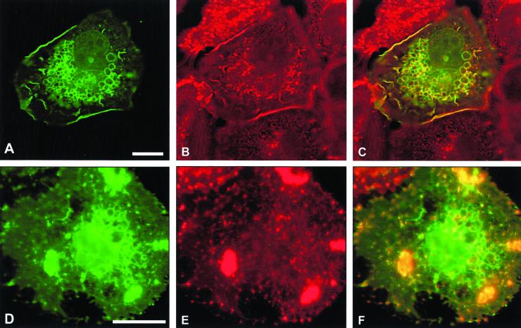
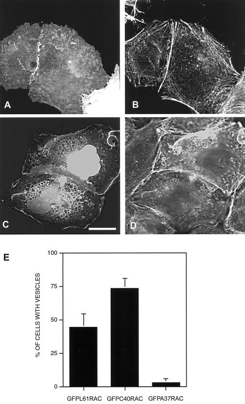
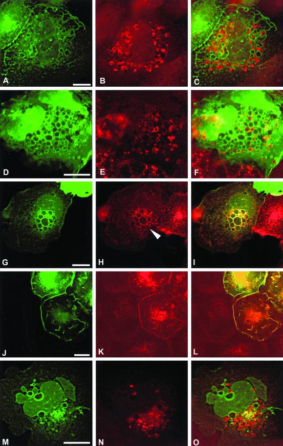
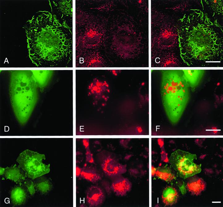
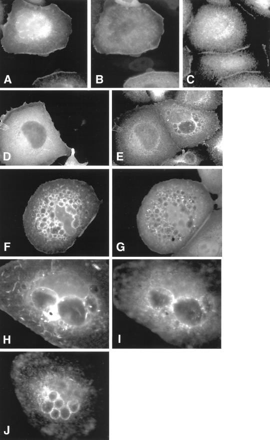
The establishment of cadherin-dependent cell-cell contacts in human epidermal keratinocytes are known to be regulated by the Rac1 small GTP-binding protein, although the mechanisms by which Rac1 participates in the assembly or disruption of cell-cell adhesion are not well understood. In this study we utilized green fluorescent protein (GFP)-tagged Rac1 expression vectors to examine the subcellular distribution of Rac1 and its effects on E-cadherin-mediated cell-cell adhesion. Microinjection of keratinocytes with constitutively active Rac1 resulted in cell spreading and disruption of cell-cell contacts. The ability of Rac1 to disrupt cell-cell adhesion was dependent on colony size, with large established colonies being resistant to the effects of active Rac1. Disruption of cell-cell contacts in small preconfluent colonies was achieved through the selective recruitment of E-cadherin-catenin complexes to the perimeter of multiple large intracellular vesicles, which were bounded by GFP-tagged L61Rac1. Similar vesicles were observed in noninjected keratinocytes when cell-cell adhesion was disrupted by removal of extracellular calcium or with the use of an E-cadherin blocking antibody. Moreover, formation of these structures in noninjected keratinocytes was dependent on endogenous Rac1 activity. Expression of GFP-tagged effector mutants of Rac1 in keratinocytes demonstrated that reorganization of the actin cytoskeleton was important for vesicle formation. Characterization of these Rac1-induced vesicles revealed that they were endosomal in nature and tightly colocalized with the transferrin receptor, a marker for recycling endosomes. Expression of GFP-L61Rac1 inhibited uptake of transferrin-biotin, suggesting that the endocytosis of E-cadherin was a clathrin-independent mechanism. This was supported by the observation that caveolin, but not clathrin, localized around these structures. Furthermore, an inhibitory form of dynamin, known to inhibit internalization of caveolae, inhibited formation of cadherin vesicles. Our data suggest that Rac1 regulates adherens junctions via clathrin independent endocytosis of E-cadherin.


Figures

Figure 1
Figure 1
Expression of constitutively active Rac1 disrupts cell–cell contacts by recruitment of E-cadherin into large intracellular vesicles. Keratinocytes cultured in normal growth medium were microinjected with cDNA constructs expressing GFP-tagged dominant negative Rac1 (GFP-N17Rac1; A–C) or GFP-tagged constitutively active Rac1 (GFP-L61Rac1; D–L). Cells were fixed and permeabilized (A–I) or fixed without permeabilization (J–L) 16 h after microinjection and stained for E-cadherin (B, E, and K) or desmoplakin-1 (H). Distribution of GFP-L61Rac1 and E-cadherin, or desmoplakin-1 was compared by merging images (C, F, I, and L). Images are from 0.4-μm sections taken 4.4 μm from basal surface of the cell. Keratinocyte colony size was <60 cells in all cases. Scale bar, 10 μm.
Figure 2
Figure 2
Vesicle formation is not a consequence of microinjection or overexpression of GFP-L61Rac1 relative to endogenous Rac1. To compare expression levels of exogenous GFP-Rac1 relative to endogenous Rac1 keratinocytes were microinjected with a cDNA construct expressing GFP-L61Rac1 (A) and costained with an antibody that recognizes Rac1 (B). Large established colonies of noninjected keratinocytes (>200 cell size) were also fixed and stained with an antibody to E-cadherin. Cells at the colony edge were photographed to demonstrate the presence of E-cadherin–positive vesicles in noninjected keratinocytes (C). Scale bar, 10 μm.
Figure 3
Figure 3
Colocalization of GFP-L61Rac1 and catenins around vesicles. Keratinocytes cultured in normal growth medium were microinjected with a cDNA construct expressing GFP-L61Rac1. Sixteen hours after injection cells were fixed, permeabilized, and stained with antibodies to α-, β- or γ-catenin (B, D, and F, respectively). GFP-L61Rac1–expressing cells were detected by GFP expression (A, C, and E). Images are from 0.4-μm sections taken 4.4 μm from basal surface of the cell. Scale bar, 10 μm.
Figure 4
Figure 4
Rac1 function is dependent on stability of cadherin-dependent contacts. Keratinocytes cultured in normal growth medium (>200 cells/colony) were microinjected with a cDNA construct expressing GFP-L61Rac1. Sixteen hours after injection cells were either maintained in normal growth medium (A and B) or switched to low-calcium medium for 6 h (C and D) and costained for E-cadherin (B and D). GFP-L61Rac1–expressing cells were visualized by GFP expression (A and C). Scale bar, 10 μm. To quantitate vesicle formation cell counts were performed on small (<60 cells/colony) or large (>200 cells/colony) colonies expressing GFP-L61Rac1 in normal growth medium (+Ca2+) or in low-calcium medium (−Ca2+). GFP-L61Rac1–expressing cells were scored positive if they contained a single large (>1 μm) vesicle. Data are mean + SD from three experiments, with a minimum of 50 injected cells counted per experiment.
Figure 5
Figure 5
Disruption of cell–cell contacts results in vesicle formation and redistribution of E-cadherin and endogenous Rac1 to the perimeter of these vesicles. Small keratinocyte colonies (<60 cells/colony) were cultured in normal medium (A and D) or in low-calcium medium to disrupt cell–cell adhesion (B and C). Alternatively cell–cell adhesion in cells cultured in normal medium was disrupted by addition of antibody to E-cadherin (HECD-1) for 12 h (D). Cells were stained with antibodies to E-cadherin (A, B, and D) or Rac1 (C). Images are from 0.4-μm sections taken 4.8 μm from basal surface of the cell. Scale bar, 10 μm.
Figure 6
Figure 6
Dominant negative Rac1 inhibits vesicle formation in cells cultured in medium containing low calcium. Keratinocytes cultured in normal growth medium were microinjected with DNA constructs expressing GFP alone (A and B) or GFP-tagged N17Rac1 (C and D). Sixteen hours after injection, cells were switched to low-calcium medium for 4 h and costained for E-cadherin (B and D). Images are from 0.4-μm sections taken 4.8 μm from basal surface of the cell. Scale bar, 10 μm.
Figure 7
Figure 7
Cycloheximide treatment does not block localization of E-cadherin to intracellular vesicles. Keratinocytes cultured in normal growth medium were treated with cycloheximide to block protein synthesis (B) or left untreated (A) and switched to low-calcium medium for 4 h to induce vesicles. Cells were fixed and stained for E-cadherin expression. Images are from 0.4-μm sections taken 4.4 μm from basal surface of the cell. Scale bar, 10 μm. To control for activity of cycloheximide¤ keratinocyte lysates were prepared from cells treated with cycloheximide (+CHX) or left untreated (−CHX) and subsequently were maintained in normal growth medium (+Ca2+) or switched to low-calcium medium (−Ca2+). Fifteen micrograms of protein from each lane was resolved by SDS-PAGE, Western blotted, and immunoblotted with antibody to E-cadherin (C, lanes 1–4) or c-myc (C, lanes 5–8).
Figure 8
Figure 8
Colocalization of actin and GFP-L61Rac1 around vesicles. Keratinocytes cultured in normal growth medium (<60 cells/colony) were microinjected with a DNA construct expressing GFP-tagged L61Rac1. Sixteen hours after injection, cells were treated with cytochalasin D for 30 min (D–F) or left untreated (A–C). Cells were fixed and stained for filamentous actin using Texas Red–conjugated phalloidin (B and E). Cells expressing the GFP-L61Rac1 construct were detected by GFP expression (A and D), and localization was compared by merging images (C and F). Images are from 0.4-μm sections taken 4.0 μm from basal surface of the cell. Scale bar, 10 μm.
Figure 9
Figure 9
Vesicle formation is an actin-dependent process. Keratinocytes cultured in normal growth medium (<60 cells/colony) were microinjected with DNA constructs expressing GFP-L61A37Rac1 (A and B) or GFPL61C40Rac1 (C and D). Sixteen hours after injection, cells were fixed and stained with Texas Red–conjugated phalloidin (B and D). Expression of the Rac1 constructs was visualized by expression of GFP (A and C). Images are from 0.4-μm sections taken 4.4 μm from basal surface of the cell. Scale bar, 10 μm. Cells counts were performed on cells microinjected with DNA constructs expressing GFP-L61Rac1, GFP-L61C40Rac1, or GFPL61A37Rac1. Injected cells expressing each construct were scored positive if they contained one or more large (>1 μm) vesicles. Data are mean + SD from three experiments, with a minimum of 50 injected cells counted per experiment.
Figure 10
Figure 10
Characterization of Rac1-induced vesicles. Keratinocytes cultured in normal growth medium were microinjected with a DNA construct expressing GFP-tagged L61Rac1. Sixteen hours after injection cells were fixed and stained for an ER marker (SERCA, B), an early endosome marker (EEA1, E), transferrin receptor (H and K), and a marker of late endosomes (CD63, N). Expression of injected constructs was visualized by expression of GFP (A, D, G, J, and M), and localization was compared by merging images (C, F, I, L, and O). Images are from 0.4-μm sections taken 4.4 μm from basal surface of the cell, except for J–L, which were taken 5.6 μm from basal surface of the cell. Scale bar, 10 μm.
Figure 11
Figure 11
Fluid phase uptake of transferrin and Texas Red dextran. Keratinocytes cultured in normal growth medium (<60 cells/colony) were microinjected with pCDNA-GFP-L61Rac1 (A, D, and G). Uptake of Texas Red dextran (E and H) and biotinylated transferrin (B) was assessed as described in MATERIALS AND METHODS. Incorporation of Texas Red into Rac-induced vesicles was monitored in live cells without fixation or permeabilization (D–F). Merged images show distribution of GFP-L61Rac1 (in green) and transferrin biotin or Texas Red biotin (in red). Scale bar, 10 μm
Figure 12
Figure 12
Rac1–induced vesicle formation is a clathrin-independent mechanism. Keratinocytes cultured in normal growth medium (<60 cells/colony) were coinjected with pCDNA-GFP-L61Rac1 (A) and a cDNA expressing a dominant negative form of dynamin I (K44A; B) and left for 16 h before fixation. Expression of injected constructs was visualized by expression of GFP (A) and staining for dynamin (B). Distribution of E-cadherin was visualized using a polyclonal pan-cadherin antibody (C). In a separate experiment, keratinocytes were microinjected with a DNA construct expressing dominant negative form of dynamin I (K44A) and cultured overnight in normal growth medium to allow expression, before being switched to low-calcium medium for 4 h. Cells were fixed and stained for dynamin to detect expression of the dominant negative dynamin construct (D) and E-cadherin using a polyclonal pan-cadherin antibody (E). To analyze distribution of clathrin and caveolin, keratinocytes microinjected with pCDNA-GFP-L61Rac1 were stained with antibodies to clathrin (F) and caveolin (H). Expression of GFP-L61Rac1 was visualized by GFP expression (G and I). Keratinocytes (<60 cells/colony) were also cultured in low-calcium medium for 4 h to induce vesicle formation, fixed, and stained with an antibody to caveolin (J).

References

    1. Akhtar N, Hudson KR, Hotchin NA. Co-localization of Rac1 and E-cadherin in human epidermal keratinocytes. Cell Adh Comm. 2000;7:465–476. - PubMed
    1. Braga VM, Del Maschio A, Machesky L, Dejana E. Regulation of cadherin function by Rho and Rac: modulation by junction maturation and cellular context. Mol Biol Cell. 1999;10:9–22. - PMC - PubMed
    1. Braga VM, Machesky LM, Hall A, Hotchin NA. The small GTPases Rho and Rac are required for the establishment of cadherin-dependent cell-cell contacts. J Cell Biol. 1997;137:1421–1431. - PMC - PubMed
    1. Bretscher MS, Aguado-Velasco C. EGF induces recycling membrane to form ruffles. Curr Biol. 1998;8:721–724. - PubMed
    1. Damke H, Baba T, van der Bliek AM, Schmid SL. Clathrin-independent pinocytosis is induced in cells overexpressing a temperature-sensitive mutant of dynamin. J Cell Biol. 1995;131:69–80. - PMC - PubMed

Publication types

MeSH terms