. 2001 Apr;12(4):919-30.
doi: 10.1091/mbc.12.4.919.

Distinct modes of macrophage recognition for apoptotic and necrotic cells are not specified exclusively by phosphatidylserine exposure

Affiliations
Free PMC article

Distinct modes of macrophage recognition for apoptotic and necrotic cells are not specified exclusively by phosphatidylserine exposure

R E Cocco et al. Mol Biol Cell. 2001 Apr.
Free PMC article

Abstract

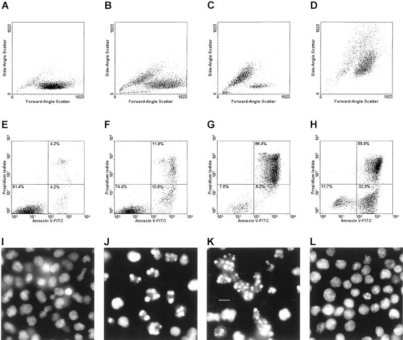
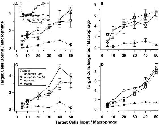
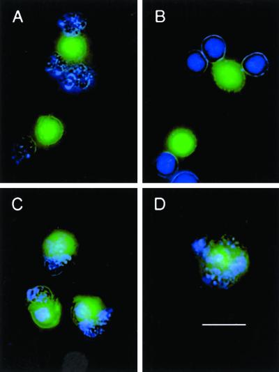
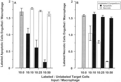
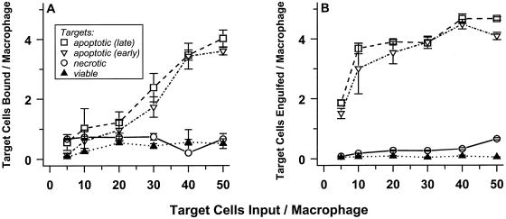
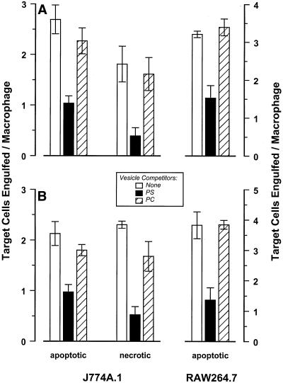
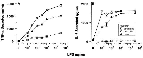
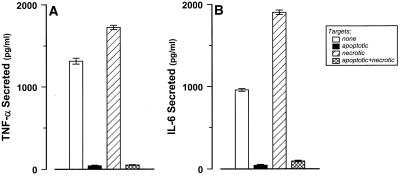
The distinction between physiological (apoptotic) and pathological (necrotic) cell deaths reflects mechanistic differences in cellular disintegration and is of functional significance with respect to the outcomes that are triggered by the cell corpses. Mechanistically, apoptotic cells die via an active and ordered pathway; necrotic deaths, conversely, are chaotic and passive. Macrophages and other phagocytic cells recognize and engulf these dead cells. This clearance is believed to reveal an innate immunity, associated with inflammation in cases of pathological but not physiological cell deaths. Using objective and quantitative measures to assess these processes, we find that macrophages bind and engulf native apoptotic and necrotic cells to similar extents and with similar kinetics. However, recognition of these two classes of dying cells occurs via distinct and noncompeting mechanisms. Phosphatidylserine, which is externalized on both apoptotic and necrotic cells, is not a specific ligand for the recognition of either one. The distinct modes of recognition for these different corpses are linked to opposing responses from engulfing macrophages. Necrotic cells, when recognized, enhance proinflammatory responses of activated macrophages, although they are not sufficient to trigger macrophage activation. In marked contrast, apoptotic cells profoundly inhibit phlogistic macrophage responses; this represents a cell-associated, dominant-acting anti-inflammatory signaling activity acquired posttranslationally during the process of physiological cell death.


Figures

Figure 1
Figure 1
Morphological discrimination of apoptotic and necrotic cell deaths. DO11.10 cells were left untreated (viable; A, E, and I) or were induced to die. To induce physiological death, cells were treated with actinomycin D (200 ng/ml) for 8 h (early apoptotic; B, F, and J) or for 18 h (late apoptotic; C, G, and K). Pathological death resulted from heating (necrotic, 55°C, 15 min; D, H, and L). Cells were analyzed for attributes of death. Changes in cellular size and granularity were evaluated cytofluorometrically with respect to forward-angle and side-angle light scatter (A–D). Membrane integrity and the accessibility of phosphatidylserine also were assessed cytofluorometrically by staining with propidium iodide (PI; Exλ = 488 nm, Emλ = 610 nm) and FITC-conjugated annexin V (Exλ = 488 nm, Emλ = 525 nm), respectively (E–H). The relative distribution within each population of viable (annexin V PI) cells and those with early (annexin V+ PI) and late (annexin V+ PI+) manifestations of death are indicated. Note that an early population of annexin V+ PI dying necrotic cells (discussed in the text) is evident in H. Chromatin condensation was visualized microscopically after staining with Hoechst 33342 (Exλ = 355 nm; Emλ = 465; I –L; bar, 5 μm).
Figure 2
Figure 2
J774A.1 macrophages selectively bind and engulf apoptotic and necrotic cells. Target cells were covalently labeled with CFDA and were induced to die physiologically (apoptotic) or pathologically (necrotic). Apoptotic death of DO11.10 cells (A and B) was triggered by treatment with actinomycin D; apoptotic S49 cells (C and D) resulted from treatment with dexamethasone. Populations of early (≤20% Trypan Blue+; ▿) and late (≥65% Trypan Blue+; □) apoptotic targets were prepared. In contrast, necrotic targets (○) were ≥80% Trypan Blue+, whereas untreated (viable; ▴) cells were <8% Trypan Blue+. Graded numbers of labeled cells were mixed with adherent murine J774A.1 macrophages in microwells. After incubation for 60 min at 4°C, unbound cells were removed by washing, and the number of target cells bound was determined by quantifying CFDA fluorescence (Exλ = 490 nm; Emλ = 525 nm; A and C). Similarly, the extent of engulfment (which may include low numbers of target cells that are bound and not engulfed) was determined after incubation for 60 min at 37°C (B and D). In the absence macrophages, binding of target cells to microwells was insignificant.
Figure 3
Figure 3
Visualization of macrophage binding and engulfment of target cells. J774A.1 macrophages were labeled (green) with 5 μM CFDA and plated in microplates with coverslip bottoms. DO11.10 cells were treated with actinomycin D for 18 h to induce apoptotic targets (A and C); necrotic targets were heat-killed (B and D). After staining (blue) with Hoechst 33342, target cells were added to macrophages at a ratio of 20:1, and cells were incubated for 60 min at 4°C (binding; A and B) and at 37°C (engulfment, as well as binding; C and D). Unbound target cells then were removed by washing; visibly bound targets were not dislodged by this procedure. The image in B illustrates that homotypic target cell association can amplify the apparent extent of binding; on average, this phenomenon distorts the magnitude of specific binding by <10%. Under these conditions, the extent to which target cells aggregated in the absence of macrophages was <3% in all cases. Images are composites of blue Hoechst staining, green CFDA labeling, and phase-contrast illumination of cell outlines. Bar, 10 μm.
Figure 4
Figure 4
Apoptotic and necrotic cells do not compete for recognition by J774A.1 macrophages. Recognition of late apoptotic and necrotic CFDA-labeled DO11.10 targets (A and B, respectively) was assessed in the presence or absence of unlabeled target competitors after incubation for 30 min at 37°C, as in Figure 2. Targets were prepared as in Figure 1. Labeled targets (2 × 10⁵ cells/well) were mixed with unstained apoptotic (▪) or necrotic (□) competitors at the indicated ratios and added to a freshly plated monolayer (2 × 10⁴ cells/well) of J774A.1 macrophages.
Figure 5
Figure 5
RAW 264.7 macrophages do not recognize necrotic cells. Viable (▴), necrotic (○), and early (▿) and late (□) apoptotic DO11.10 targets were presented to RAW 264.7 macrophages, and binding (A) and engulfment (B) were assessed as in Figure 2.
Figure 6
Figure 6
Phosphatidylserine is not a specific ligand for apoptotic cell recognition. J774A.1 and Raw 264.7 macrophages were plated in microwells and incubated in the absence (A) or in the presence of LPS (0.1 μg/ml, B) for 3 h. DO11.10 targets (as in Figure 1) were added to the macrophage monolayers at a ratio of 10:1. Phosphatidylserine (PS; ▪]) or phosphatidylcholine (PC; square with diagonal line) liposomes were added as indicated to a final phospholipid concentration of 25 μM. Target cell interactions with macrophages in the presence or absence (□) of these liposomes were assessed after incubation at 37°C for 60 min.
Figure 7
Figure 7
Engulfed apoptotic cells inhibit proinflammatory cytokine release. J774A.1 macrophages were incubated at 37°C without or with LPS at the indicated concentrations for 2 h before the addition of late apoptotic (□) or necrotic (○) DO11.10 targets (prepared as in Figure 1; target to macrophage ratio of 20:1) or without added targets (▴). Culture supernatants were collected 4 h after target addition, and levels of secreted TNF-α and IL-6 were quantified. Under these conditions, target cells themselves produced no detectable levels of cytokines (<10 pg/ml TNF-α and < 15 pg/ml IL-6).
Figure 8
Figure 8
Inhibition of cytokine release from macrophages exerted by apoptotic cells is a dominant effect. J774A.1 macrophages were incubated with LPS (100 ng/ml) for 2 h DO11.10 targets (as in Figure 1) were added to wells subsequently (at the indicated target to macrophage ratios): late apoptotic (▪; 10:1), necrotic (▨; 10:1), apoptotic and necrotic (⊠; 10 + 10:1). Secreted TNF-α and IL-6 levels were quantified 4 h after target addition or in the absence of any added targets (□).

References

    1. Albert ML, Pearce SFA, Francisco LM, Sauter B, Roy P, Silverstein RL, Bhardwaj N. Immature dendritic cells phagocytose apoptotic cells via aVβ5 and CD36, and cross-present antigens to cytotoxic T lymphocytes. J Exp Med. 1998;188:1359–1368. - PMC - PubMed
    1. Bellingan GJ, Caldwell H, Howie SEM, Dransfield I, Haslett C. In vivo fate of the inflammatory macrophage during the resolution of inflammation: inflammatory macrophages do not die locally, but emigrate to the draining lymph nodes. J Immunol. 1996;157:2577–2585. - PubMed
    1. Bratton DL, Fadok VA, Richter DA, Kailey JM, Guthrie LA, Henson PM. Appearance of phosphatidylserine on apoptotic cells requires calcium-mediated nonspecific flip-flop and is enhanced by loss of the aminophospholipid translocase. J Biol Chem. 1997;272:26159–26165. - PubMed
    1. Cecconi F, Alvarez-Bolado G, Meyer BI, Roth KA, Gruss P. Apaf1 (CED-4 homolog) regulates programmed cell death in mammalian development. Cell. 1998;94:727–737. - PubMed
    1. Chang M-K, Bergmark C, Laurila A, Hörkkö S, Han K-H, Friedman P, Dennis EA, Witztum JL. Monoclonal antibodies against oxidized low-density lipoprotein bind to apoptotic cells and inhibit their phagocytosis by elicited macrophages: evidence that oxidation-specific epitopes mediate macrophage recognition. Proc Natl Acad Sci USA. 1999;96:6353–6358. - PMC - PubMed

Publication types

LinkOut - more resources