. 2001 Apr;12(4):957-69.
doi: 10.1091/mbc.12.4.957.

Deletion of yeast p24 genes activates the unfolded protein response

Affiliations
Free PMC article

Deletion of yeast p24 genes activates the unfolded protein response

W J Belden et al. Mol Biol Cell. 2001 Apr.
Free PMC article

Abstract

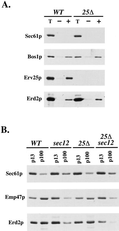
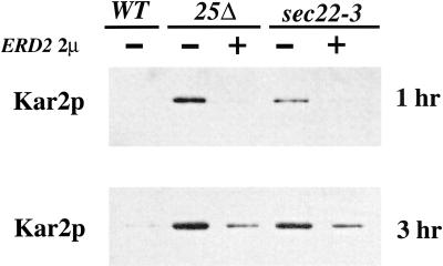
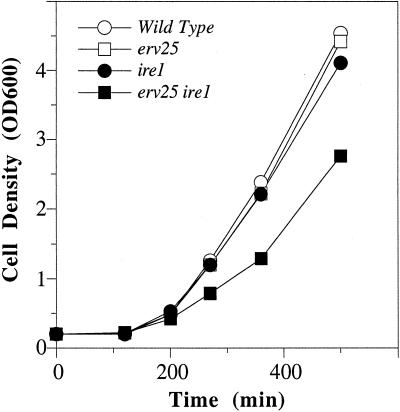
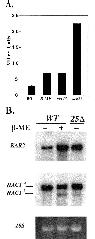
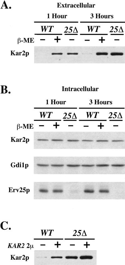
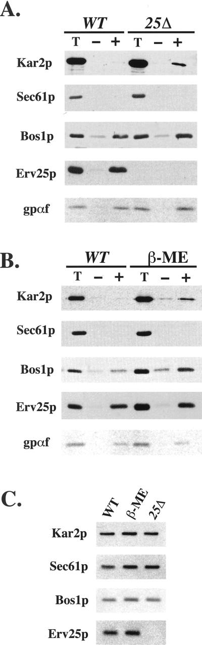
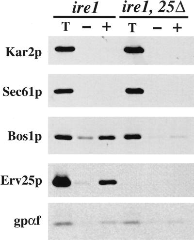
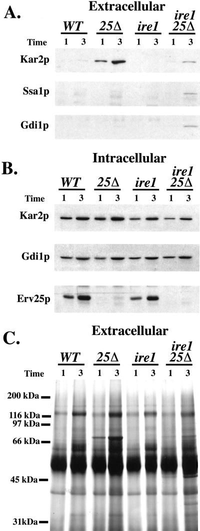
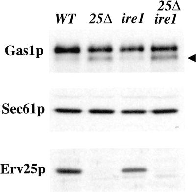
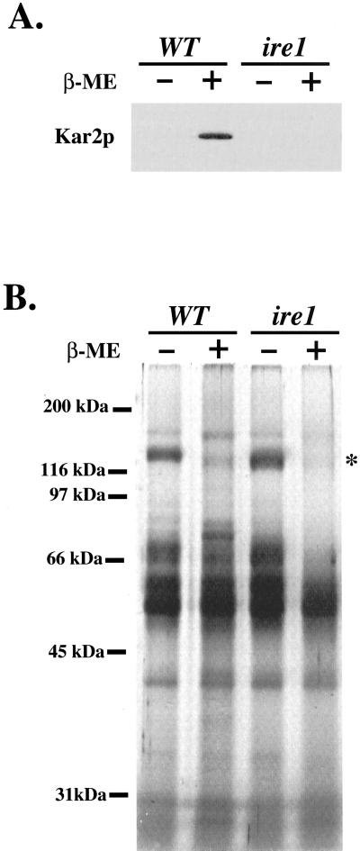
Yeast cells lacking a functional p24 complex accumulate a subset of secretory proteins in the endoplasmic reticulum (ER) and increase the extracellular secretion of HDEL-containing ER residents such as Kar2p/BiP. We report that a loss of p24 function causes activation of the unfolded protein response (UPR) and leads to increased KAR2 expression. The HDEL receptor (Erd2p) is functional and traffics in p24 deletion strains as in wild-type strains, however the capacity of the retrieval pathway is exceeded. Other conditions that activate the UPR and elevate KAR2 expression also lead to extracellular secretion of Kar2p. Using an in vitro assay that reconstitutes budding from the ER, we detect elevated levels of Kar2p in ER-derived vesicles from p24 deletion strains and from wild-type strains with an activated UPR. Silencing the UPR by IRE1 deletion diminished Kar2p secretion under these conditions. We suggest that activation of the UPR plays a major role in extracellular secretion of Kar2p.


Figures

Figure 1
Figure 1
Erd2p transport between the ER and Golgi compartments is independent of the p24 complex. (A) Reconstituted COPII budding reactions were performed on ER membranes isolated from CBY398 (WT) and CBY419 (erv25Δ) strains that express an epitope-tagged version of Erd2p. Lanes labeled T represent one-tenth of the total membranes used in a budding reaction, minus (−) lanes indicate the amount of vesicles formed in the absence of the purified COPII components, and plus (+) lanes indicate vesicles produced when COPII proteins are added. Total membranes and budded vesicles were collected by centrifugation, resolved on a polyacrylamide gel, and immunoblotted for indicated proteins. (B) ER and Golgi membrane fractions from CBY398 (WT), CBY548 (sec12), CBY549 (erv25Δ), and CBY550 (sec12, erv25Δ) strains that express the myc-tagged version of Erd2p. Cells were shifted to 37OC for 1 h before harvesting and lysis. ER membranes were pelleted by a medium-speed centrifugation (p13) and Golgi membranes were collected by a high-speed centrifugation (p100). Proteins contained in these fractions were monitored by immunoblot analysis.
Figure 2
Figure 2
Overexpression of ERD2 partially suppresses the secretion of Kar2p in an erv25Δ deletion strain. The amount of extracellular Kar2p secreted after 1 and 3 h in erv25Δ and sec22-3 strains with and without an ERD2 2 μ plasmid. Proteins contained in the cell culture supernatant were concentrated by TCA precipitation and Kar2p was detected by immunoblot.
Figure 3
Figure 3
Growth rate is decreased in an erv25Δ ire1 double mutant. The optical density at 600 nm was determined for FY834 (WT), CBY114 (erv25), CBY427 (ire1), and CBY425 (ire1 erv25) in rich media at 30OC and plotted as a function of time.
Figure 4
Figure 4
(A) Deletion of ERV25 causes activation of the UPR. β-Galctosidase assays were performed on CBY635 (WT), CBY635 treated with 15 mM β-mercaptoethanol (β-ME) for 60 min, CBY637 (erv25Δ), and CBY748 (sec22-3). Activity is given in Miller Units and represents the average of seven independent determinations with SE. There is a significant difference (p < 0.0001) between the wild-type and erv25Δ strain. (B) Northern blot analysis of a FY834 (WT) and CBY114 (25Δ) with (+) or without (−) 15 mM β-mercaptoethanol (β-ME) treatment. Blots were probed for KAR2 and HAC1 messages with 32P-labeled probes. Spliced (HAC1i) and unspliced (HAC1U) were observed. Ethidium stained 18 S rRNA serves as a loading control.
Figure 5
Figure 5
Activation of the UPR or overproduction of Kar2p increases extracellular Kar2p secretion. (A) Amount of secreted Kar2p from FY834 (WT) cells treated with (+) and without (−) 15 mM β-mercaptoethanol (β-ME) compared with CBY114 (25Δ). (B) Intracellular levels of Kar2p, Gdi1p, and Erv25p from equivalent amounts of cells (based on OD600) grown as in A. (C) Extracellular Kar2p from FY834 (WT) and CBY114 (25Δ) that were transformed (+) or not (−) with a 2 μ KAR2 plasmid. Proteins contained in the cell culture supernatant were concentrated by TCA precipitation and Kar2p was detected by immunoblot.
Figure 6
Figure 6
Deletion of ERV25 or activation of the UPR increases the amount of Kar2p in COPII vesicles. (A) Reconstituted COPII budding reactions were performed on ER membranes isolated from FY834 (WT) and CBY114 (25Δ). Lanes labeled T represent one-tenth of the total membranes used in a budding reaction, minus (−) lanes indicate the amount of vesicles formed in the absence of the purified COPII components, and plus (+) lanes indicate vesicles produced when COPII proteins are added. Total membranes and budded vesicles were collected by centrifugation, resolved on polyacrylamide gels, and immunoblotted for indicated proteins. The Kar2p detected represents protease protected material (see MATERIALS AND METHODS) and [35S]-glyco-pro-α-factor (gpαf) was monitored by fluorography. (B) As in A except ER membranes were prepared from FY834 (WT) or FY834 treated with 15 mM β-mercaptoethanol (β-ME), to activate the UPR. (C) Equal amounts of ER membranes (microsomes) blotted with indicated markers to compare levels of Kar2p.
Figure 7
Figure 7
COPII budding is decreased in an erv25Δ ireΔ double mutant. (A) Reconstituted COPII budding reactions were performed as in Figure 6 except ER membranes were prepared form CBY427 (ire1 Δ) and CBY425 (ire1 Δ, 25Δ).
Figure 8
Figure 8
Deletion of IRE1 prevents Kar2p secretion from an erv25Δ strain. (A) Amount of extracellular Kar2p in cultures from FY834 (WT), CBY114 (erv25 Δ), CBY427 (ire1 Δ), and CBY425 (erv25 Δ ire1 Δ) after 1 and 3 h. Proteins contained in the cell culture supernatant were concentrated by TCA precipitation and Kar2p was detected by immunoblot. (B) Intracellular levels of Kar2p, Gdi1p, and Erv25p from equal volumes of cell cultures grown as in A. (C) The same samples of precipitated proteins as in A were resolved on 12.5% SDS-PAGE and silver stained.
Figure 9
Figure 9
Immunoblot analysis of wild-type and deletion strains. Whole cell membranes prepared from FY834 (WT), CBY114 (erv25 Δ), CBY427 (ire1 Δ), and CBY425 (erv25 Δ ire1 Δ) strains after 3 h. Membrane proteins were resolved on polyacrylamide gels and immunoblotted for Gas1p, Sec61p (loading control), and Erv25p. The arrowhead indicates the ER form of Gas1p.
Figure 10
Figure 10
Deletion of IRE1 prevents Kar2p secretion from cells treated with β-mercaptoethanol. (A) Amount of extracellular Kar2p from FY834 (WT) or CBY427 (ire1 Δ) cells treated with (+) and without (−) 15 mM β-mercaptoethanol (β-ME) for 3 h. Proteins contained in the cell culture supernatant were concentrated by TCA precipitation and Kar2p was detected by immunoblot. (B) The same samples of precipitated proteins as in A were resolved on 12.5% SDS-PAGE and silver stained.

References

    1. Ausubel FM, Brent R, Kingston RE, Moore DD, Seidman JG, Smith JA, Struhl K. Current Protocols in Molecular Biology. New York: John Wiley & Sons; 1997.
    1. Bagnat M, Keranen S, Shevchenko A, Shevchenko A, Simons K. Lipid rafts function in biosynthetic delivery of proteins to the cell surface in yeast. Proc Natl Acad Sci USA. 2000;97:3254–3259. - PMC - PubMed
    1. Baker D, Hicke L, Rexach M, Schleyer M, Schekman R. Reconstitution of SEC gene product-dependent intercompartmental protein transport. Cell. 1988;54:335–344. - PubMed
    1. Balch WE, McCaffery JM, Plutner H, Farquhar MG. Vesicular stomatitis virus glycoprotein is sorted and concentrated during export from the endoplasmic reticulum. Cell. 1994;76:841–852. - PubMed
    1. Barlowe C, Orci L, Yeung T, Hosobuchi M, Hamamoto S, Salama N, Rexach MF, Ravazzsola M, Amherdt M, Schekman R. COPII: a membrane coat formed by Sec proteins that drive vesicle budding from the endoplasmic reticulum. Cell. 1994;77:895–907. - PubMed

Publication types

MeSH terms

LinkOut - more resources