. 2001 Apr;12(4):971-80.
doi: 10.1091/mbc.12.4.971.

Cell cycle-dependent changes in microtubule dynamics in living cells expressing green fluorescent protein-alpha tubulin

Affiliations
Free PMC article

Cell cycle-dependent changes in microtubule dynamics in living cells expressing green fluorescent protein-alpha tubulin

N M Rusan et al. Mol Biol Cell. 2001 Apr.
Free PMC article

Abstract

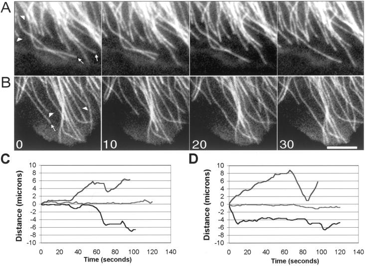
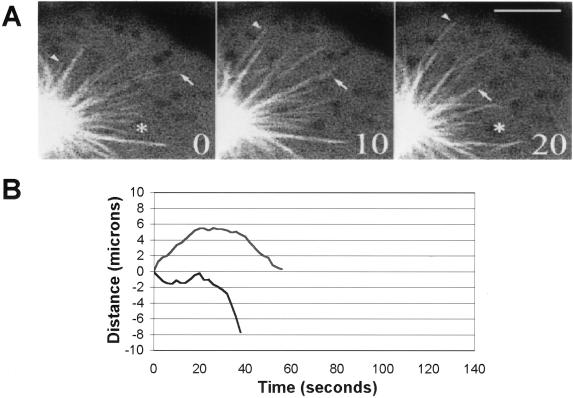
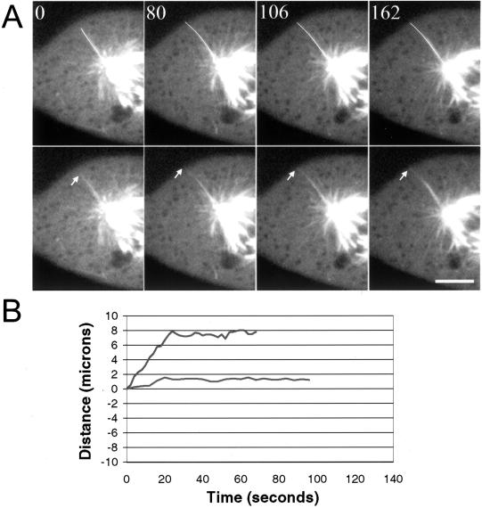
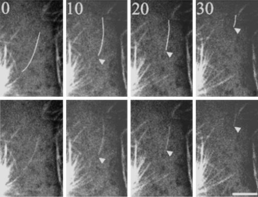
LLCPK-1 cells were transfected with a green fluorescent protein (GFP)-alpha tubulin construct and a cell line permanently expressing GFP-alpha tubulin was established (LLCPK-1alpha). The mitotic index and doubling time for LLCPK-1alpha were not significantly different from parental cells. Quantitative immunoblotting showed that 17% of the tubulin in LLCPK-1alpha cells was GFP-tubulin; the level of unlabeled tubulin was reduced to 82% of that in parental cells. The parameters of microtubule dynamic instability were compared for interphase LLCPK-1alpha and parental cells injected with rhodamine-labeled tubulin. Dynamic instability was very similar in the two cases, demonstrating that LLCPK-1alpha cells are a useful tool for analysis of microtubule dynamics throughout the cell cycle. Comparison of astral microtubule behavior in mitosis with microtubule behavior in interphase demonstrated that the frequency of catastrophe increased twofold and that the frequency of rescue decreased nearly fourfold in mitotic compared with interphase cells. The percentage of time that microtubules spent in an attenuated state, or pause, was also dramatically reduced, from 73.5% in interphase to 11.4% in mitosis. The rates of microtubule elongation and rapid shortening were not changed; overall dynamicity increased 3.6-fold in mitosis. Microtubule release from the centrosome and a subset of differentially stable astral microtubules were also observed. The results provide the first quantitative measurements of mitotic microtubule dynamics in mammalian cells.


Figures

Figure 1
Figure 1
Image sequences of microtubule dynamic behavior in living LLCPK-1 cells injected with rhodamine tubulin (A) and in LLCPK-1α cells expressing GFP-tubulin (B). Images were collected at 2-s intervals and are shown at 10-s intervals. Arrows indicate shortening events; arrowheads indicate growth events. Bar, 5 μm. Life history plots showing the dynamic behavior of individual microtubules in LLCPK-1 cells injected with rhodamine tubulin (C) and LLCPK-1α cells expressing GFP-tubulin (D). The life history plots shown in C and D are from different cells than shown in A and B. Most microtubules in these cells displayed periods of growth, shortening, and pause (upper and lower traces in C and D); a subset of microtubules was differentially stable (middle traces, C and D). Only cells in which >50% of the trackable microtubules were dynamic were included in the data set; for all cells analyzed, all trackable microtubules in the cell were included in the analysis.
Figure 2
Figure 2
Microtubule behavior in mitotic cells. (A) Dynamic behavior of astral microtubules in LLCPK-1α cells; images shown were collected at 10-s intervals by using the Bio-Rad confocal microscope. The arrowhead shows a microtubule that grows out from the aster center; the arrow shows a shortening microtubule. The asterisk marks the same pixel location in the first and last images. Bar, 5 μm. (B) Life history plots of mitotic microtubles in LLCPK-1α cells. Each line on the graph represents the excursions made by an individual microtubule plus-end during the observation period. The majority of microtubules either grew out from the aster, followed by a catastrophe transition (upper trace), or were in the field of view at the start of the image sequence and subsequently underwent a catastrophe and disassembled out of the field of view (lower trace). The life history plots shown in B are from different cells than shown in A.
Figure 3
Figure 3
Differentially stabilized astral microtubules in mitotic cells. (A) Image sequence showing an astral microtubule that grows out to the cell edge and then pauses for the remainder of the sequence. The microtubule end is marked by the arrow. In the upper panels, the microtubule has been colored white to aid in visualization. Images were acquired using the Ultraview confocal microscope at 2-s intervals; Bar, 5 μm. (B) Life history plots of the dynamic behavior of individual stable, astral microtubules. The microtubule shown in the upper trace grew toward the cell cortex where it was relatively stable for the remainder of the movie sequence; the microtubule in the lower trace was inactive for the entire sequence. The life history plots shown in B are from different cells than shown in A.
Figure 4
Figure 4
Astral microtubules are released from the centrosome in mitotic cells. In the upper panels the released microtubule has been colored white to aid in visualization. In this sequence, a microtubule is released and appears to move away from the aster center; the microtubule end was not clear before the image labeled t = 0. The minus-end most likely moved into the field of view by transport or disassembly following release; no evidence for a severing event was detected. Time is given in seconds in each frame. Bar, 5 μm.

References

    1. Andersen SSL. Balanced regulation of microtubule dynamics during the cell cycle: a contemporary view. BioEssays. 1999;21:53–60. - PubMed
    1. Belmont LD, Hyman AA, Sawin KE, Mitchison TJ. Real-time visualization of cell cycle dependent changes in microtubule dynamics in cytoplasmic extracts. Cell. 1990;62:579–589. - PubMed
    1. Berrueta L, Kraeft S-K, Tirnauer JS, Schuyler SC, Chen LB, Hill DE, Pellman D, Bierer BE. The adenomatous polyposis coli-binding protein EB1 is associated with cytoplasmic and spindle microtubules. Proc Natl Acad Sci USA. 1998;95:10596–10601. - PMC - PubMed
    1. Busson S, Dujardin D, Moreau A, Dompierre J, DeMay JR. Dynein and dynactin are localized to astral microtubules and at cortical sites in mitotic epithelial cells. Curr Biol. 1998;8:541–544. - PubMed
    1. Carminati J, Stearns T. Microtubules orient the mitotic spindle in yeast through dynein-dependent interactions with the cell cortex. J Cell Biol. 1997;138:629–641. - PMC - PubMed

Publication types

LinkOut - more resources