. 2001 Apr 17;20(8):1840-9.
doi: 10.1093/emboj/20.8.1840.

Expression of microbial virulence proteins in Saccharomyces cerevisiae models mammalian infection

Affiliations

Expression of microbial virulence proteins in Saccharomyces cerevisiae models mammalian infection

C F Lesser et al. EMBO J. .

Abstract

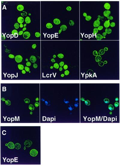
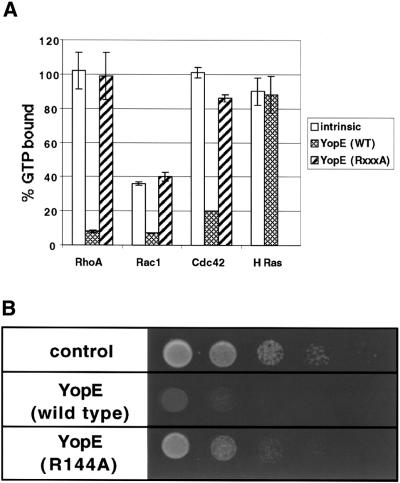
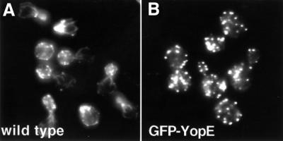
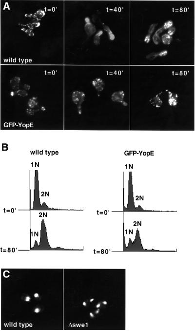
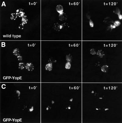
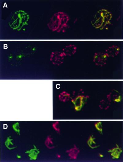
Bacterial virulence proteins that are translocated into eukaryotic cells were expressed in Saccharomyces cerevisiae to model human infection. The subcellular localization patterns of these proteins in yeast paralleled those previously observed during mammalian infection, including localization to the nucleus and plasma membrane. Localization of Salmonella SspA in yeast provided the first evidence that SspA interacts with actin in living cells. In many cases, expression of the bacterial virulence proteins conferred genetically exploitable growth phenotypes. In this way, Yersinia YopE toxicity was demonstrated to be linked to its Rho GTPase activating protein activity. YopE blocked polarization of the yeast cytoskeleton and cell cycle progression, while SspA altered polarity and inhibited depolymerization of the actin cytoskeleton. These activities are consistent with previously proposed or demonstrated effects on higher eukaryotes and provide new insights into the roles of these proteins in pathogenesis: SspA in directing formation of membrane ruffles and YopE in arresting cell division. Thus, study of bacterial virulence proteins in yeast is a powerful system to determine functions of these proteins, probe eukaryotic cellular processes and model mammalian infection.


Figures

None
Fig. 1. Immunofluorescence microscopy of yeast expressing Y.pseudotuberculosis virulence proteins. Yeast (W303a) carrying high-copy-number plasmids encoding galactose-inducible GFP fusion genes to each of the indicated bacterial virulence proteins were visualized 1 h (A and B) or 3 h (C) after the addition of galactose. (B) Yeast were fixed in ethanol and stained with DAPI to visualize DNA. GFP fused to the virulence protein (green) is shown in the first panel, DNA (blue) is shown in the second panel and the third panel (YopM/DAPI) represents the first two panels merged together such that the turquoise features represent co-localization of GFP–YopM (green) and DNA (blue).
None
Fig. 2. Yeast growth is inhibited by expression of YpkA or YopE. Yeast (W303a) carrying high-copy-number plasmids encoding galactose-inducible GFP fusion genes to the indicated bacterial protein were grown overnight in non-inducing selective synthetic media containing raffinose as a carbon source. Cultures were then normalized to OD600 = 1 and serial 10-fold dilutions were spotted onto selective media plates containing raffinose (1%) and galactose (2%). Photographs were taken after 2 days of growth at 30°C.
None
Fig. 3. YopE toxicity is dependent on RhoGAP activity. (A) Purified His-tagged YopE and mutant (R144A) YopE-R144A (5.5 pmol) were incubated with G-proteins (GST–Rac1, GST–RhoA, GST–Cdc42 and GST–H-Ras) (5.5 pmol) that were pre-loaded with [γ-32P]GTP. (His6-tagged YopE-R144A was not incubated with GST–H-Ras.) GTPase activity was assayed after 5 min. Values represent the mean ± SD of three separate experiments. (B) Growth assays (as described in Figure 2) were conducted on cells transformed with a high-copy-number plasmid encoding galactose-inducible mutant GFP-yopE (R144A) and compared to cells transformed with empty vector or wild-type GAL10-GFP-yopE.
None
Fig. 4. YopE expression disrupts polarity of the actin cytoskeleton. Wild-type (A) and W303::GAL10-GFP-yopE (B) yeast were grown in non-inducing selective liquid media containing raffinose (2%) and sorbitol (1 M) to OD600 = 0.3–0.6. Galactose was added to these asynchronous cultures to a final concentration of 2%. Yeast were then harvested after 2 h, fixed with formaldehyde and stained with rhodamine-labeled phalloidin to visualize the actin cytoskeleton.
None
Fig. 5. YopE blocks bud formation and arrests cells at the morphogenesis checkpoint. Yeast were grown in non-inducing YEP (W303 strains) or selective (S288C strains) liquid media containing raffinose (2%) and sorbitol (1 M) to OD600 = 0.3–0.6. The cultures were then synchronized with α-factor for 2.5 h such that they arrested at G1 and formed mating projections (schmoos). Galactose (2%) was added to the synchronized cultures. After 1 h, α-factor was removed and cells were harvested at 40 min intervals. (A) Wild-type and W303::GAL10-GFP-yopE yeast were fixed with formaldehyde and stained with rhodamine-labeled phalloidin to visualize the actin cytoskeleton. (B) Wild-type and W303::GAL10-GFP-yopE yeast were fixed with ethanol and stained with PI, and subjected to flow cytometry analysis. 1N and 2N represent the DNA content of the two peaks. (C) Wild-type or Δswe1 yeast (S288Ca background) carrying galactose-inducible GFP-yopE encoded on a low-copy plasmid (pCFL112) were fixed with ethanol and stained with PI to visualize the DNA/nuclei.
None
Fig. 6. YopE blocks cytokinesis and formation of actin rings. Wild-type and W303::GAL10-GFP-yopE yeast were grown in non-inducing YEP liquid media containing raffinose (2%) and sorbitol (1 M) to OD600 = 0.3–0.6. The cultures were then synchronized with nocodazole for 2 h such that they arrested as large budded cells at the spindle checkpoint. Galactose (2%) was added to the synchronized cultures. After 1 h, nocodazole was removed and cells were harvested at 60 min intervals. Cells were fixed with formaldehyde and stained with rhodamine-labeled phalloidin to visualize the actin cytoskeleton (A and B) and with DAPI (C) to visualize DNA/nuclei.
None
Fig. 7. GFP–SspA co-localizes with and reorganizes the actin cytoskeleton. Each panel represents immunofluorescent images of yeast (W303a) carrying plasmids encoding galactose-inducible GFP–sspA. Yeast were fixed with formaldehyde at the designated time points and stained with rhodamine-labeled phalloidin to visualize actin. In (A, B and D), GFP–SspA (green) is shown in the first panel, actin (red) is shown in the second panel and the third panel represents the first two panels merged together such that yellow features indicate co-localization of GFP–SspA (green) and actin (red). In (C), each panel represents merged GFP–SspA (green) and actin (red) images such that the yellow features represent co-localization of GFP–SspA and actin. Six hours after induction, GFP–SspA expressed from a high-copy plasmid (pCFL170) co-localized with all of the actin in the yeast and disrupted normal cell polarity. (B) One hour after induction, GFP–SspA, expressed from a single copy of the integrated fusion gene (YCL170), co-localized with a small subset of yeast cortical patches. (C) Three hours after induction, GFP–SspA, expressed from a single copy of the integrated fusion gene (YCL170) co-localized with actin cables that appear to originate from GFP–SspA-containing cortical patches. (D) Twelve hours after induction of GFP–SspA expression from a low-copy plasmid (pCFL172), only the actin cytoskeleton that co-localized with GFP–SspA was resistant to a 60 min exposure to LatA (200 µM).

Similar articles

Cited by

References

    1. Adams A.E., Shen,W., Lin,C.S., Leavitt,J. and Matsudaira,P. (1995) Isoform-specific complementation of the yeast sac6 null mutation by human fimbrin. Mol. Cell. Biol., 15, 69–75. - PMC - PubMed
    1. Ayscough K.R., Stryker,J., Pokala,N., Sanders,M., Crews,P. and Drubin,D.G. (1997) High rates of actin filament turnover in budding yeast and roles for actin in establishment and maintenance of cell polarity revealed using the actin inhibitor latrunculin-A. J. Cell Biol., 137, 399–416. - PMC - PubMed
    1. Belmont L.D. and Drubin,D.G. (1998) The yeast V159N actin mutant reveals roles for actin dynamics in vivo. J. Cell Biol., 142, 1289–1299. - PMC - PubMed
    1. Black D.S. and Bliska,J.B. (2000) The RhoGAP activity of the Yersinia pseudotuberculosis cytotoxin YopE is required for antiphagocytic function and virulence. Mol. Microbiol., 37, 515–527. - PubMed
    1. Bourdet-Sicard R., Egile,C., Sansonetti,P.J. and Tran Van Nhieu,G. (2000) Diversion of cytoskeletal processes by Shigella during invasion of epithelial cells. Microbes Infect., 2, 813–819. - PubMed

Publication types

MeSH terms