. 2001 Apr 17;20(8):1921-30.
doi: 10.1093/emboj/20.8.1921.

mAKAP assembles a protein kinase A/PDE4 phosphodiesterase cAMP signaling module

Affiliations

mAKAP assembles a protein kinase A/PDE4 phosphodiesterase cAMP signaling module

K L Dodge et al. EMBO J. .

Abstract

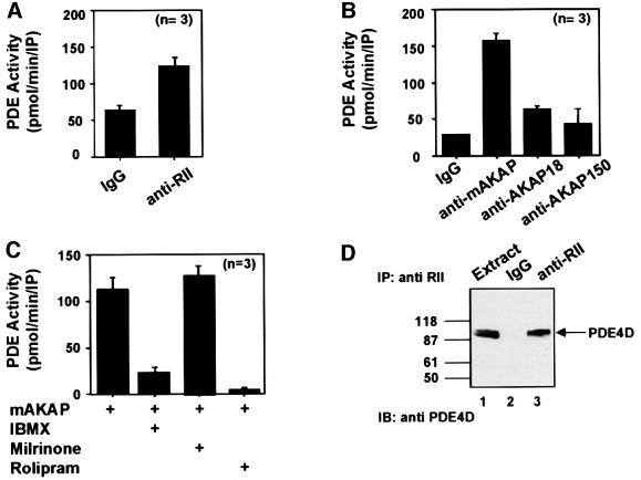
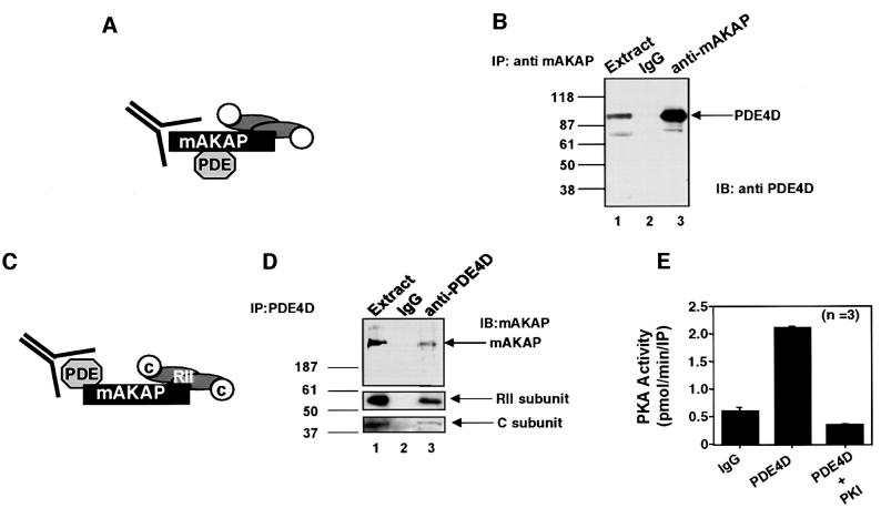
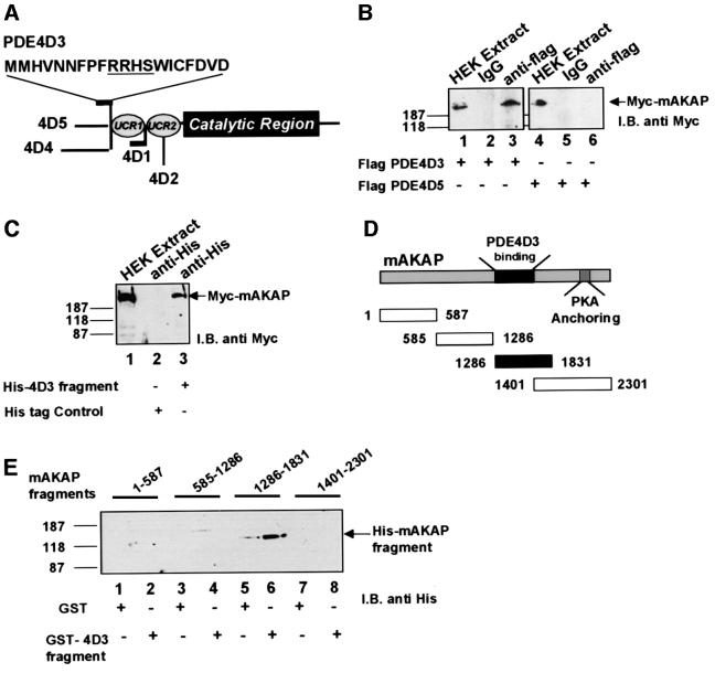
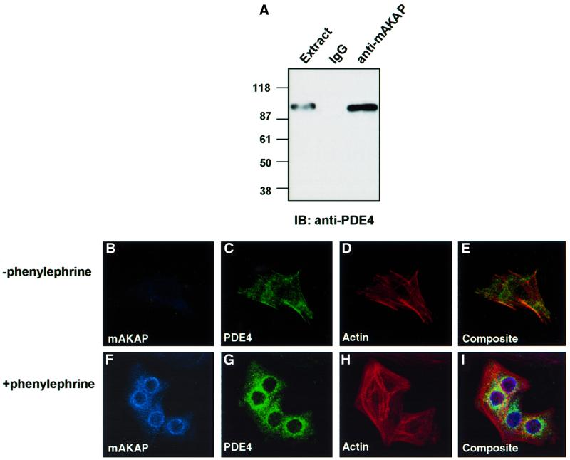
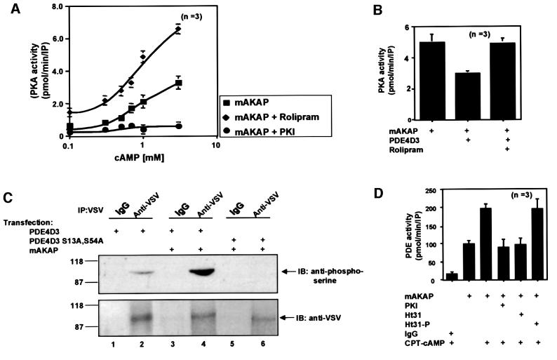
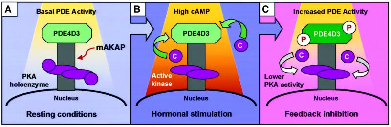
Spatiotemporal regulation of protein kinase A (PKA) activity involves the manipulation of compartmentalized cAMP pools. Now we demonstrate that the muscle-selective A-kinase anchoring protein, mAKAP, maintains a cAMP signaling module, including PKA and the rolipram-inhibited cAMP-specific phosphodiesterase (PDE4D3) in heart tissues. Functional analyses indicate that tonic PDE4D3 activity reduces the activity of the anchored PKA holoenzyme, whereas kinase activation stimulates mAKAP-associated phosphodiesterase activity. Disruption of PKA- mAKAP interaction prevents this enhancement of PDE4D3 activity, suggesting that the proximity of both enzymes in the mAKAP signaling complex forms a negative feedback loop to restore basal cAMP levels.


Figures

None
Fig. 1. Type 4 PDE activity co-purifies with mAKAP. (A) Immune complexes were isolated from rat heart extracts using antibodies against the RII subunit of cAMP-dependent protein kinase or control IgG serum. Co-precipitating PDE activity [pmol/min/immunoprecipitation (IP)] was measured using [3H]cAMP as a substrate. Data are presented as the average of three independent experiments. (B) Immune complexes were isolated from rat heart extracts using antibodies against mAKAP, AKAP18 or control IgG and from brain extracts using antibodies against AKAP150. The source of the antibody is indicated below each column. Co-precipitating PDE activity (pmol/min/IP) was measured as described above. Data presented are the average of three independent experiments. (C) mAKAP immune complexes were treated with PDE inhibitors (indicated below each column) and PDE activity was measured as described above. Data presented are the average of three independent experiments. (D) Immunoprecipitations were performed from rat heart extracts using antibodies against the RII subunit of PKA or IgG control. The resulting immune complexes were separated by electrophoresis on a 7.5% SDS gradient polyacrylamide gel and electrotransferred to nitrocellulose membranes. Detection of PDE4D in the extract (lane 1), IgG control (lane 2) or the RII immunoprecipitation (lane 3) was by immunoblot analysis using a monoclonal antibody against the PDE4D family. Detection of signals was by chemiluminescence. Molecular weight markers are indicated. The migration of PDE4D is indicated.
None
Fig. 2. Biochemical characterization of the mAKAP signaling complex. The mAKAP signaling complex was analyzed by a series of complementary biochemical approaches. (A) A schematic diagram depicting the isolation of the mAKAP immune complexes. (B) Immunoprecipitations were performed from rat heart extracts using antibodies against rat mAKAP or control IgG. The resulting immune complexes were separated by electrophoresis on a 7.5% SDS–polyacrylamide gel and electrotransferred to nitrocellulose membranes. Immunoblot analyses using monoclonal antibodies against the PDE4D family were used to identify the PDE in heart extracts (lane 1) and immunoprecipitations with the IgG control (lane 2) or anti-mAKAP antisera (lane 3). Detection of signals was by chemiluminescence. Molecular weight markers are indicated. The migration of PDE4D is indicated. (C) A schematic diagram depicting immunoprecipitation of PDE4D and associated proteins. (D) Immune complexes were isolated from rat heart extracts (lane 1) using goat polyclonal antibodies against PDE4D isoforms (lane 3) or control goat IgG (lane 2). Immune complexes were separated by electrophoresis on a 7.5% SDS–polyacrylamide gel and electrotransferred to nitrocellulose membranes. The filter was subjected to immunoblot analysis using rabbit polyclonal antibodies against rat mAKAP (top panel), RII regulatory subunit (middle panel) and catalytic subunit of PKA (bottom panel). Detection of signals was by chemiluminescence. Molecular weight markers and the migration position of mAKAP are indicated. (E) PKA activity in PDE4D immune complexes was measured using the heptapeptide Kemptide as a substrate. PKA specific activity (pmol/min/IP) was measured from PDE4D immune complexes, the IgG control and in the presence of the PKI 5–24 peptide, a specific inhibitor of PKA. The source of the sample is indicated below each lane. The accumulated data from three experiments are presented.
None
Fig. 3. PDE4D3 directly binds a central region of mAKAP. Mapping studies were performed to determine which PDE4D isoform interacts with mAKAP. (A) A schematic diagram of the PDE4D gene family highlights the conserved catalytic core and upstream conserved regions (UCR). The locations of divergent sequences in each isoform are indicated. The unique first 15 residues of PDE4D3 are presented using the one letter amino acid code. (B) Recombinant Flag-tagged PDE4D3 or Flag-tagged PDE4D5 was expressed in HEK293 cells. Anti-Flag antibodies or control IgG were used to isolate immune complexes. The precipitates were separated by gel electrophoresis on a 7.5% SDS–polyacrylamide gel and electrotransferred to nitrocellulose membranes. Co-precipitation of epitope-tagged mAKAP from cell extracts expressing PDE4D3 (lanes 1–3) or PDE4D5 (lanes 4–6) was assessed by immunoblotting (I.B.) using monoclonal antibodies against the myc tag. Detection of the signal was by chemiluminescence. Molecular weight markers and the migration position of Myc-mAKAP are indicated. (C) A His-tagged fusion protein encompassing the unique region of PDE4D3 or a His-tagged control fragment was co-expressed with mAKAP in HEK293 cells. Anti-His-tag antisera were used to isolate immune complexes. The presence of mAKAP was assessed by immunoblotting using anti-Myc monoclonal antibodies in HEK 293 cell extracts (lane 1) and in immune complexes from cells expressing His-PDE4D3 fragment (lane 3) or control fragment (lane 2). Detection of the signal was by chemiluminescence. Molecular weight markers and the migration position of Myc-mAKAP are indicated. A family of His-tagged-mAKAP fragments encompassing distinct regions of the anchoring protein were expressed in Escherichia coli. (D) A schematic diagram shows the size of each mAKAP fragment and its location within the linear sequence of the anchoring protein. The first and last amino acids of each fragment are indicated. The region of the anchoring protein that interacts with the PDE (closed box) and the binding site for PKA are highlighted. (E) A GST fusion protein including the unique N-terminal sequence of PDE4D3 was used to screen the purified mAKAP fragments by in vitro pull-down assay (lanes 2, 4, 6 and 8). Control experiments were performed with GST alone (lanes 1, 3, 5 and 7). The isolated materials were separated by gel electrophoresis on a 7.5% SDS–polyacrylamide gel and electrotransferred to nitrocellulose membranes. Isolation of mAKAP fragments was assessed by immunoblotting (I.B.) using monoclonal antibodies against the His tag. Detection of the signal was by chemiluminescence. Molecular weight markers are indicated.
None
Fig. 4. Recruitment of mAKAP and PDE4D3 to the perinuclear regions of RNVs upon induction of hypertrophy. Expression of mAKAP is upregulated upon the induction of hypertrophy in RNV, resulting in accumulation of the anchoring protein at perinuclear regions (Kapiloff et al., 1999). (A) As a prelude to immunocytochemical analysis, control experiments confirmed that a monoclonal antibody against PDE4 family members recognized the PDE that co-precipitated with mAKAP from cardiomyocyte extracts. Immunoprecipitations were performed using antibodies against rat mAKAP or control IgG. The resulting immune complexes were separated by electrophoresis on a 7.5% SDS–polyacrylamide gel and electrotransferred to nitrocellulose membranes. Immunoblot analysis using a monoclonal antibody that recognizes the PDE4 family was used to identify the PDE in immunoprecipitations with anti-mAKAP antisera or the IgG control (indicated above each lane). This antibody was subsequently used in immunofluorescence experiments. Dissociated RNVs were plated on coverslips and grown in the absence (upper panels) or presence (lower panels) of phenylephrine (100 µM) for 24 h at 37°C. Ventriculocytes were fixed by incubation in 3.7% formaldehyde for 10 min at room temperature, permeablized with 0.3% Triton X-100, and subjected to labeling with primary antibodies against mAKAP (1:500 dilution) and PDE4 family(1:500 dilution) for 16 h at 4°C. The intracellular location of mAKAP (blue; B and F) was detected with Cy5-labeled secondary antibodies (1:100 dilution), PDE4 (green; C and G) was detected with fluorescein-labeled secondary antibodies (1:100 dilution) and the actin cytoskeleton (red; D and H) was detected with Texas red–phalloidin. Immunofluorescent detection of each signal was performed by confocal microscopy on a BioRad 1024 UV/Vis confocal microscope. Composite images are presented in (E) and (I).
None
Fig. 5. mAKAP assembles a PDE/PKA negative feedback loop. The functional consequences of PDE4D3 localization through association with mAKAP were assessed on PKA activity. (A) The mAKAP signaling complex was immunoprecipitated from rat heart extracts using antibodies against the mouse anchoring protein. PKA activity (squares) was measured over a range of cAMP concentrations (0.1–10 mM) as described in Materials and methods, upon inhibition of PDE4D3 by rolipram (diamonds) and in the presence of the PKI 5–24 peptide (circles). The specific activity of the PKA catalytic subunit is presented as pmol/min/IP. The averaged data from three experiments are presented. (B) Recombinant myc-tagged mAKAP was expressed in HEK293 cells in the absence or presence of PDE-4D3 (indicated below each lane). Anti-Myc antibodies were used to isolate immune complexes and PKA activity was measured in the presence of 1 mM cAMP. The specific activity of the PKA catalytic subunit is presented as pmol/min/IP. The averaged data from three experiments are presented. Rolipram (10 µM) was used to selectively inhibit PDE activity. (C) Expression vectors encoding VSV-tagged PDE4D3 (lanes 1–4) or the phosphorylation site mutant VSV-tagged PDE4D3 S13A,S54A (lanes 5 and 6) were co-expressed with myc-tagged mAKAP in HEK293 (lanes 2–6). The transfected plasmids are indicated above each lane. The cells were incubated with 1 mM CPT-cAMP for 15 min before immunoprecipitation with anti-VSV antibodies or control IgG. The immune complexes were separated by gel electrophoresis on a 7.5% SDS–polyacrylamide gel and electrotransferred to nitrocellulose membranes. Phosphorylation of PDE4D3 was assessed by immunoblotting (IB) using a polyclonal phosphoserine-specific antibody (top panel). Equal loading of PDE4D3 was confirmed by reprobing the blot with VSV-specific monoclonal antibody (bottom panel). Detection of the signals was by chemiluminescence. Molecular weight markers and the migration position of PDE are indicated. This figure is representative of three independent experiments. (D) In reciprocal experiments, the functional consequences of PKA anchoring were assessed on PDE4D3 activity. The mAKAP signaling complex was immunoprecipitated from rat heart extracts using antibodies against the rat anchoring protein. After a 16 h incubation, the immunoprecipitates were incubated in the presence or absence of CPT-cAMP for 30 min (indicated below). PDE4 activity was then measured under conditions where PKA activity was inhibited (PKI), in the presence of a PKA anchoring inhibitor peptide (Ht31) or a control peptide (Ht31p). PDE activity was presented as pmol/min/IP. The data averaged from three experiments are presented.
None
Fig. 6. mAKAP maintains an anchored PKA/PDE4D3 negative feedback loop. The cartoon depicts the proposed feedback inhibition of anchored PKA by mAKAP-associated PDE4DE3 activity. (A) Under basal conditions, the PKA holoenzyme is dormant and the localized constitutively active PDE maintains cAMP concentrations below an activation threshold for the kinase. (B) Upon hormonal stimulation, the increased flow of second messenger overcomes the rate of PDE-mediated cAMP degradation. This promotes release of the PKA catalytic (C) subunit. (C) PKA phosphorylation of mAKAP-associated PDE4D3 stimulates PDE activity, thereby driving cAMP levels back to basal. This favors reformation of the PKA holoenzyme.

Similar articles

Cited by

References

    1. Beard M.B., O’Connell,J.C., Bolger,G.B. and Houslay,M.D. (1999) The unique N-terminal domain of the cAMP phosphodiesterase PDE4D4 allows for interaction with specific SH3 domains. FEBS Lett., 460, 173–177. - PubMed
    1. Beard M.B., Olsen,A.E., Jones,R.E., Erdogan,S., Houslay,M.D. and Bolger,G.B. (2000) UCR1 and UCR2 domains unique to the cAMP-specific phosphodiesterase family form a discrete module via electrostatic interactions. J. Biol. Chem., 275, 10349–10358. - PubMed
    1. Beavo J.A. (1995) Cyclic nucleotide phosphodiesterases: functional implications of multiple isoforms. Physiol. Rev., 75, 725–748. - PubMed
    1. Beavo J.A., Bechtel,P.J. and Krebs,E.G. (1974) Preparation of homogenous cyclic AMP-dependent protein kinase(s) and its subunits from rabbit skeletal muscle. Methods Enzymol., 38, 299–308. - PubMed
    1. Beavo J.A., Conti,M. and Heaslip,R.J. (1994) Multiple cyclic nucleotide phosphodiesterases. Mol. Pharmacol., 46, 399–405. - PubMed

Publication types

Substances