. 2001 Apr 17;20(8):2028-40.
doi: 10.1093/emboj/20.8.2028.

MOT1-catalyzed TBP-DNA disruption: uncoupling DNA conformational change and role of upstream DNA

Affiliations

MOT1-catalyzed TBP-DNA disruption: uncoupling DNA conformational change and role of upstream DNA

R P Darst et al. EMBO J. .

Abstract

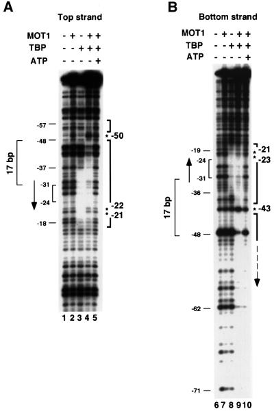
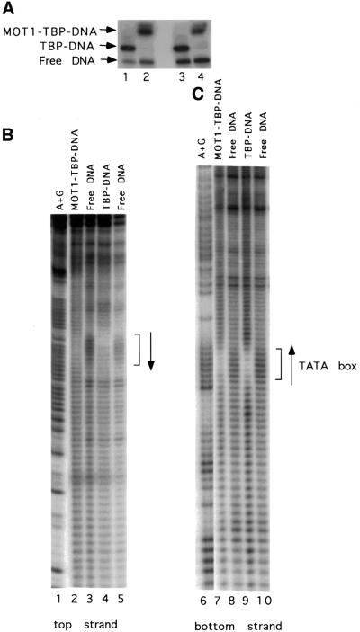
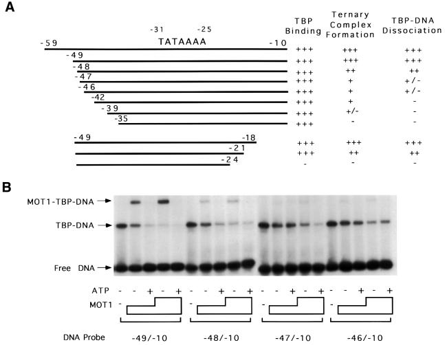
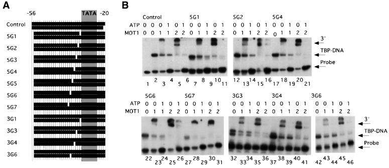
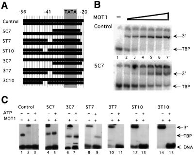
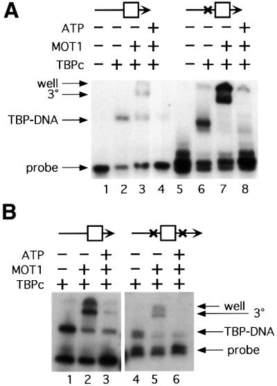
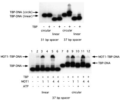
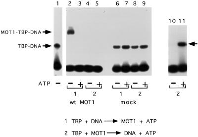
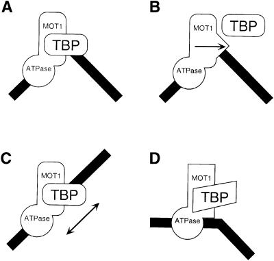
SNF2/SWI2-related ATPases employ ATP hydrolysis to disrupt protein-DNA interactions, but how ATP hydrolysis is coupled to disruption is not understood. Here we examine the mechanism of action of MOT1, a yeast SNF2/SWI2-related ATPase that uses ATP hydrolysis to remove TATA binding protein (TBP) from DNA. MOT1 function requires a 17 bp DNA 'handle' upstream of the TATA box, which must be double stranded. Remarkably, MOT1-catalyzed disruption of TBP-DNA does not appear to require DNA strand separation, DNA bending or twisting of the DNA helix. Thus, TBP-DNA disruption is accomplished in a reaction apparently not driven by a change in DNA structure. MOT1 action is supported by DNA templates in which the handle is connected to the TATA box via single-stranded DNA, indicating that the upstream duplex DNA can be conformationally uncoupled from the TATA box. Combining these results with proposed similarities between SNF2/SWI2 ATPases and helicases, we suggest that MOT1 uses ATP hydrolysis to translocate along the handle and thereby disrupt interactions between TBP and DNA.


Figures

None
Fig. 1. DNase I footprinting of TBP–DNA and MOT1–TBP–DNA complexes. A radiolabeled 110 bp DNA probe (∼0.15 nM) containing a consensus TATA sequence labeled on either the top (A) or the bottom strand (B) was incubated with 5 nM TBP and/or 20 nM MOT1 and 10 µM ATP as indicated. The reactions were treated with DNase I and reaction products were resolved on a denaturing high resolution polyacrylamide gel and visualized by autoradiography as described in Materials and methods.
None
Fig. 2. Identification of nucleosides required for TBP and MOT1 interaction with DNA by hydroxyl radical treatment. The same radiolabeled DNA probes as used in Figure 1 were pre-treated with hydroxyl radical to remove random nucleosides. The collection of damaged DNAs (∼3 nM) was then incubated with 5 nM TBP and/or 20 nM MOT1 as indicated, and the free DNA and protein–DNA complexes were resolved by non-denaturing PAGE (A). The indicated bands were then excised and resolved on high resolution denaturing polyacrylamide gels to identify the nucleosides critical for interaction with TBP and MOT1 (B and C). Note the depletion of DNA molecules missing TATA box nucleosides in the reactions containing TBP and TBP plus MOT1.
None
Fig. 3. Formation of the MOT1–TBP–DNA complex requires 17 bp of DNA upstream of the TATA box. Radiolabeled DNA probes (0.15 nM) of the lengths indicated were incubated with 5 nM TBP, TBP plus 1 or 4 U (∼6 or 24 nM) of MOT1, and TBP plus MOT1 and 10 µM ATP. Protein–DNA complexes were detected by non-denaturing gel electrophoresis. The results are summarized in (A); electrophoretic mobility shift results using probes that bracket the boundary between functional and non-functional DNAs are shown in (B).
None
Fig. 4. DNA containing single base pair gaps is competent for MOT1 binding and ATP-driven disruption of TBP–DNA. (A) Diagram of DNA constructs. The control construct was based upon the adenovirus major late promoter (numbers indicate the position in the promoter), with most AT base pairs being changed to GC for the cross-linking experiment (below; see Table I for oligonucleotide sequences). Gray shading indicates the position of the sequence TATAAAAG. Eleven constructs with 1 bp gaps in the upstream DNA were made. Label was placed on the TATA-less upstream strand, so no shift is seen unless the construct anneals fully. (B) Gel mobility shift results using fully duplex control DNA (lanes 1–5) and nine of the constructs made. TBP core domain was used at 5 nM. The reaction in lane 1 contained no TBP. ATP, added where indicated, was used at 5 µM. Units of MOT1 were added as indicated. One unit of MOT1 is estimated to be 6 nM. The DNA concentration is ∼0.05 nM. Positions of ternary complex (‘3°’), TBP–DNA complex (‘TBP–DNA’) and unbound DNA probe (‘Probe’) are indicated. Results with 5G3 and 5G5 (not shown) are identical. On construct 3G3, two discrete TBP–DNA complexes were detected; the reason for this is unknown but was specific for a template with a gap at this position.
None
Fig. 5. Effects of large gaps in the upstream DNA on MOT1 binding and ATP-driven disruption of TBP–DNA. (A) Diagram of DNA constructs. The control is the same as in Figure 4; other constructs are derived from control, missing sequences at the positions indicated. Upstream segments are labeled. (B) Gel mobility shift results using fully duplex control DNA and the 5C7 template, which has an 18 base single-stranded tail. The positions of the MOT1–TBP–DNA complex (‘3°’) and TBP–DNA complex (‘TBP’) are indicated; the free DNA band is not shown. Reactions in lanes 1 contained no MOT1, 1 U of MOT1 (∼6 nM) was added to the reactions in lane 2 and the amount of MOT1 was doubled in successive lanes. (C) Gel mobility shift results obtained with each of the DNA constructs. Conditions were as described for Figure 4B, except that 4 U of MOT1 (∼24 nM) were used in each MOT1-containing reaction. Constructs 5C7 and 3C7 support ternary complex but MOT1-catalyzed disruption is severely impaired. Positions of ternary complex (‘3°’), TBP–DNA complex (‘TBP’) and free DNA probe (‘DNA’) are indicated. In (B) and (C), DNA was present at 0.15 nM and TBP was added to 5 nM concentration.
None
Fig. 6. Psoralen cross-links flanking the TATA box do not impair MOT1 function. (A) Singly cross-linked DNA compared with control. The psoralen cross-linking site is indicated (X), and the TATA box is schematized by the square. Where indicated, 5 nM TBP core domain, 1 U of MOT1 (∼6 nM) and 5 µM ATP were incubated with radiolabeled DNA probes (∼0.15 nM) constructed as described in Materials and methods. Results in lanes 1–4 are from reactions using duplex DNA with no cross-links. Lanes 5–8 contain DNA probe with a single psoralen cross-link between the upstream DNA handle and the TATA box. The positions of the free DNA, TBP–DNA complexes (‘TBP–DNA’) and MOT1–TBP–DNA complex (‘3°’) are indicated. ‘Well’ indicates the top of the gel. (B) Doubly cross-linked DNA compared with control. The procedure was the same as for (A).
None
Fig. 7. MOT1 disrupts TBP–DNA complexes formed on minicircle DNA. Gel shift experiments were performed with radiolabeled minicircle DNA (∼0.15 nM) or the identical linear sequence incubated with TBP (top panel) or TBP ± MOT1 ± ATP as indicated (bottom panel). Two sets of probes were used: circular and linear DNAs with a 31 bp spacer between the TATA box and phased A tracts, and circular and linear probes with a 37 bp spacer between the TATA box and phased A tracts. In the minicircle constructs, the 31 bp spacer pre-bends the DNA towards the minor groove of the TATA box (inhibiting TBP binding), whereas the minicircle with the 37 bp spacer pre-bends the TATA box towards the major groove, greatly enhancing the binding of TBP to this probe. The protein–DNA complexes are indicated. Reactions contained 5 nM TBP, 10 µM ATP and ∼1 or 4 U (6 or 24 nM) of MOT1 as indicated. In the top panel, note that a very small amount of residual circular DNA co-migrates with TBP–DNA complexes formed on the linear probes.
None
Fig. 8. The MOT1–TBP complex is not competent to bind DNA, but TBP is released from MOT1 in the presence of ATP. Gel mobility shift results obtained with a 40mer DNA probe that contains the upstream handle (lanes 1–9) and a 30mer DNA probe that does not support MOT1 function (lanes 10 and 11). Lane 1 shows the position of the TBP–DNA complex (also indicated by the leftward arrow adjacent to lane 11). TBP and DNA or TBP and MOT1 were pre-incubated for 20 min followed by the addition of MOT1 or DNA ± ATP for 5 min as indicated in the reaction schemes labeled 1 and 2. The reactions in lanes 2–5 and 10 and 11 contained wild-type MOT1, whereas the reactions in lanes 6–9 contained eluate obtained from a mock affinity purification using extract in which MOT1 was not epitope tagged. Note that pre-incubation of TBP and MOT1 prevents formation of protein–DNA complexes, whether or not ATP is present in the reaction (lanes 4 and 5); the effect depends on MOT1 in the reaction (lanes 8 and 9), and TBP binding to DNA can be recovered in the presence of ATP if a DNA probe is used that sequesters TBP from MOT1 action (lane 11). Reactions contained 5 nM TBP, 10 µM ATP and ∼30 nM MOT1 as indicated.
None
Fig. 9. Model for MOT1-catalyzed ATP-dependent disruption of TBP–DNA. A cartoon of the MOT1–TBP–DNA ternary complex is shown in (A). Three mechanisms are possible. MOT1 could be anchored to the DNA handle and ATP hydrolysis is used to drive a wedge to dislodge TBP (B). Alternatively, ATP hydrolysis powers translocation of MOT1 along DNA to disrupt the association of TBP with DNA (C). In these cases, duplex DNA in the handle is employed as a track to direct movement of MOT1 or a domain of MOT1. In (C), movement of MOT1 could be in either direction: movement of MOT1 to the left would cause TBP dissociation by ‘pulling’, whereas movement to the right would cause disruption of TBP–DNA by ‘pushing’. See text for discussion. (D) The upstream DNA handle might function as an allosteric activator for MOT1, which is required for MOT1 to effect a change in TBP conformation or a local change in TATA DNA structure.

Similar articles

Cited by

References

    1. Adamkewicz J.I., Mueller,C.G.F., Hansen,K.E., Prud’homme,W.A. and Thorner,J. (2000) Purification and enzymic properties of Mot1 ATPase, a regulator of basal transcription in the yeast Saccharomyces cerevisiae. J. Biol. Chem., 275, 21158–21168. - PubMed
    1. Auble D.T. and Hahn,S. (1993) An ATP-dependent inhibitor of TBP binding to DNA. Genes Dev., 7, 844–856. - PubMed
    1. Auble D.T. and Steggerda,S.M. (1999) Testing for DNA tracking by MOT1, a SNF2/SWI2 protein family member. Mol. Cell. Biol., 19, 412–423. - PMC - PubMed
    1. Auble D.T., Hansen,K.E., Mueller,C.G.F., Lane,W.S., Thorner,J. and Hahn,S. (1994) Mot1, a global repressor of RNA polymerase II transcription, inhibits TBP binding to DNA by an ATP-dependent mechanism. Genes Dev., 8, 1920–1934. - PubMed
    1. Auble D.T., Wang,D., Post,K.W. and Hahn,S. (1997) Molecular analysis of the SNF2/SWI2 protein family member MOT1, an ATP-driven enzyme that dissociates TATA-binding protein from DNA. Mol. Cell. Biol., 17, 4842–4851. - PMC - PubMed

Publication types

MeSH terms