. 2001 Apr 24;98(9):5007-12.
doi: 10.1073/pnas.071372498. Epub 2001 Apr 10.

Visualization of unwinding activity of duplex RNA by DbpA, a DEAD box helicase, at single-molecule resolution by atomic force microscopy

Affiliations

Visualization of unwinding activity of duplex RNA by DbpA, a DEAD box helicase, at single-molecule resolution by atomic force microscopy

Arnon Henn et al. Proc Natl Acad Sci U S A. .

Abstract

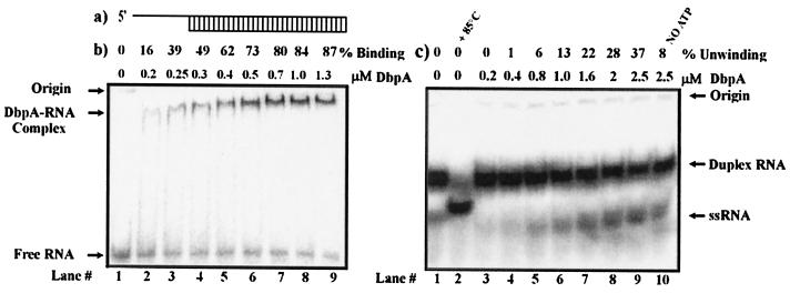
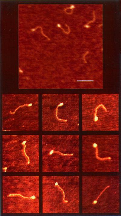
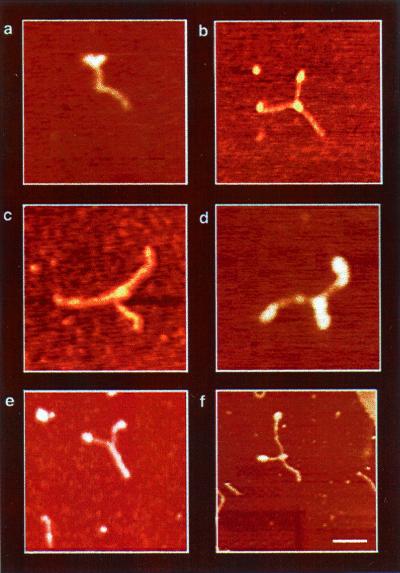
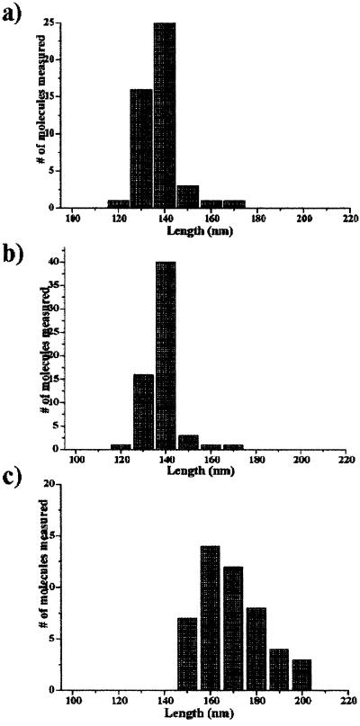
The Escherichia coli protein DbpA is unique in its subclass of DEAD box RNA helicases, because it possesses ATPase-specific activity toward the peptidyl transferase center in 23S rRNA. Although its remarkable ATPase activity had been well defined toward various substrates, its RNA helicase activity remained to be characterized. Herein, we show by using biochemical assays and atomic force microscopy that DbpA exhibits ATP-stimulated unwinding activity of RNA duplex regardless of its primary sequence. This work presents an attempt to investigate the action of DEAD box proteins by a single-molecule visualization methodology. Our atomic force microscopy images enabled us to observe directly the unwinding reaction of a DEAD box helicase on long stretches of double-stranded RNA. Specifically, we could differentiate between the binding of DbpA to RNA in the absence of ATP and the formation of a Y-shaped intermediate after its progression through double-stranded RNA in the presence of ATP. Recent studies have questioned the designation of DbpA, in particular, and DEAD box proteins in general as RNA helicases. However, accumulated evidence and the results reported herein suggest that these proteins are indeed helicases that resemble in many aspects the DNA helicases.


Figures

Figure 1
Figure 1
Binding and unwinding activity of DbpA with short dsRNA. (a) Schematic of the short dsRNA substrate used for the following assays. (b) Gel retardation assay of DbpA and dsRNA substrate. Binding reactions were performed in a 20-μl reaction volume in the presence of 5 pmol of 32P-labeled dsRNA and increasing amounts of DbpA. The binding percentages are indicated. Free RNA, RNA-DbpA complexes, and the origins of the wells in the gel are indicated on the left. (c) Unwinding of dsRNA by DbpA. Lane 1 represents free dsRNA. Lane 2 shows the ssRNA that resulted after 3 min of incubation at 85°C. Lanes 3–10 show the unwinding reaction at a given protein concentration. The differences in the position of ssRNA on the gel between lane 2 and lanes 3–10 are caused by differences in ionic strength of the free dsRNA used for control and the product of the enzymatic reaction.
Figure 2
Figure 2
AFM images of free dsRNA. (a) Schematic of the RNA construct used for the AFM imaging containing two 5′ (≈50-nt) single-stranded overhangs at both ends of the molecule. The double-stranded central region (480 bp) was generated by hybridization of the complementary regions of both strands. (b) AFM images of long dsRNA in air on mica. (Bar = 100 nm.) The rod-shaped molecules possess a mean end-to-end contour length of 134.5 ± 4 nm, which is consistent with an A form dsRNA.
Figure 3
Figure 3
AFM images of DbpA–RNA complexes. (Bar = 100 nm.) Globular structures at the end of the RNA molecule represent protein signals. The mean end-to-end contour length is 135 ± 4 nm, which is consistent with the free dsRNA. Visualization of protein signals at the end of the RNA molecule indicates that DbpA requires an ssRNA or moderate fork junction for binding before performing the unwinding activity.
Figure 4
Figure 4
AFM images of DbpA–RNA complexes in the presence of ATP (DbpA–RNA–ATP). The Y structure, observed in all images, represents the most dominant intermediate trapped during unwinding catalysis. It comprises two single ssRNA regions at the arms and dsRNA at the trunk of the Y. Protein signals were observed as globular structures at the fork junction (a--d and f) and on the tips of the Y-shaped arms (a, b, d, and f). Because of the structure of the RNA construct, two proteins can sometimes be seen entering the dsRNA region from opposite sides, as shown in d. On the other hand, e shows a Y-shaped molecule without a protein signal at the fork junction. The formation of this complex may result from substrate reannealing, or alternatively, the central protein may be washed away during sample preparations. The measured end-to-end contour length of the Y-shaped molecules shows a significant elongation (see Fig. 5c) because of the ssRNA formation during unwinding catalysis. In addition, the variation in the amount of unwinding among the Y-shaped complexes are snapshots representing the distribution of structural conformations during catalysis.
Figure 5
Figure 5
Statistical data analysis of AFM images. (a) Histogram of free dsRNA (Fig. 2a). The mean end-to-end length distribution is 134 ± 4 nm, which is consistent with A form dsRNA. (b) Same analysis of the prebound DbpA–RNA complex. The mean end-to-end contour length is similar to that shown in a. (c) Histogram of the Y-shaped contour length of the trapped DbpA–RNA–ATP complexes during unwinding catalysis. Comparison of the mean end-to-end length of the Y-shaped complexes to the free RNA and RNA + DbpA (a and b) reveals a significant shift to a longer value, which results from ssRNA formation.

References

    1. Linder P, Lasko P F, Ashburner M, Leroy P, Nielsen P J, Nishi K, Schnier J, Slonimski P P. Nature (London) 1989;337:121–122. - PubMed
    1. de la Cruz J, Kressler D, Linder P. Trends Biochem Sci. 1999;24:192–198. - PubMed
    1. Gorbalenya A E, Koonin E V, Donchenko A P, Blinov V M. Nature (London) 1988;333:22. (lett.). - PubMed
    1. Egelman E H. J Struct Biol. 1998;124:123–128. - PubMed
    1. Luking A, Stahl U, Schmidt U. Crit Rev Biochem Mol Biol. 1998;33:259–296. - PubMed

Publication types

MeSH terms

LinkOut - more resources