

Similarities between the DNA replication initiators of Gram-negative bacteria plasmids (RepA) and eukaryotes (Orc4p)/archaea (Cdc6p)
- PMID: 11296251
- PMCID: PMC33142
- DOI: 10.1073/pnas.081079298
Similarities between the DNA replication initiators of Gram-negative bacteria plasmids (RepA) and eukaryotes (Orc4p)/archaea (Cdc6p)
Abstract
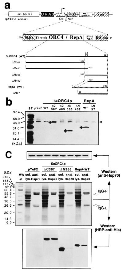
The proteins responsible for the initiation of DNA replication are thought to be essentially unrelated in bacteria and archaea/eukaryotes. Here we show that RepA, the initiator from the Pseudomonas plasmid pPS10, and the C-terminal domain of ScOrc4p, a subunit of Saccharomyces cerevisiae (Sc) origin recognition complex (ORC), share sequence similarities. Based on biochemical and spectroscopic evidence, these similarities include common structural elements, such as a winged-helix domain and a leucine-zipper dimerization motif. We have also found that ScOrc4p, as previously described for RepA-type initiators, interacts with chaperones of the Hsp70 family both in vitro and in vivo, most probably to regulate the assembly of active ORC. In evolutionary terms, our results are compatible with the recruitment of the same protein module for initiation of DNA replication by the ancestors of present-day Gram-negative bacteria plasmids, archaea, and eukaryotes.
Figures


Similar articles
-
Twenty years of the pPS10 replicon: insights on the molecular mechanism for the activation of DNA replication in iteron-containing bacterial plasmids.Plasmid. 2004 Sep;52(2):69-83. doi: 10.1016/j.plasmid.2004.06.002. Plasmid. 2004. PMID: 15336485 Review.
-
Protein domains and conformational changes in the activation of RepA, a DNA replication initiator.EMBO J. 1998 Aug 3;17(15):4511-26. doi: 10.1093/emboj/17.15.4511. EMBO J. 1998. PMID: 9687517 Free PMC article.
-
A conformational switch between transcriptional repression and replication initiation in the RepA dimerization domain.Nat Struct Biol. 2003 Jul;10(7):565-71. doi: 10.1038/nsb937. Nat Struct Biol. 2003. PMID: 12766757
-
Mechanistic aspects of DnaA-RepA interaction as revealed by yeast forward and reverse two-hybrid analysis.EMBO J. 2001 Aug 15;20(16):4577-87. doi: 10.1093/emboj/20.16.4577. EMBO J. 2001. PMID: 11500384 Free PMC article.
-
Common domains in the initiators of DNA replication in Bacteria, Archaea and Eukarya: combined structural, functional and phylogenetic perspectives.FEMS Microbiol Rev. 2003 Jan;26(5):533-54. doi: 10.1111/j.1574-6976.2003.tb00629.x. FEMS Microbiol Rev. 2003. PMID: 12586394 Review.
Cited by
-
Hsp70 Isoforms Are Essential for the Formation of Kaposi's Sarcoma-Associated Herpesvirus Replication and Transcription Compartments.PLoS Pathog. 2015 Nov 20;11(11):e1005274. doi: 10.1371/journal.ppat.1005274. eCollection 2015 Nov. PLoS Pathog. 2015. PMID: 26587836 Free PMC article.
-
Defining a novel domain that provides an essential contribution to site-specific interaction of Rep protein with DNA.Nucleic Acids Res. 2021 Apr 6;49(6):3394-3408. doi: 10.1093/nar/gkab113. Nucleic Acids Res. 2021. PMID: 33660784 Free PMC article.
-
Global Distribution and Diversity of Prevalent Sewage Water Plasmidomes.mSystems. 2022 Oct 26;7(5):e0019122. doi: 10.1128/msystems.00191-22. Epub 2022 Sep 7. mSystems. 2022. PMID: 36069451 Free PMC article.
-
Characterization of a cryptic plasmid pSM429 and its application for heterologous expression in psychrophilic Pseudoalteromonas.Microb Cell Fact. 2011 May 5;10:30. doi: 10.1186/1475-2859-10-30. Microb Cell Fact. 2011. PMID: 21542941 Free PMC article.
-
Extrachromosomal element capture and the evolution of multiple replication origins in archaeal chromosomes.Proc Natl Acad Sci U S A. 2007 Apr 3;104(14):5806-11. doi: 10.1073/pnas.0700206104. Epub 2007 Mar 28. Proc Natl Acad Sci U S A. 2007. PMID: 17392430 Free PMC article.
References
-
- Orgel L E. Trends Biochem Sci. 1998;23:491–495. - PubMed
-
- Messer W, Weigel C. In: Escherichia coli and Salmonella: Cellular and Molecular Biology. Neidhardt F C, Curtiss R III, Ingraham J L, Lin E C C, Low K B, Magasanik B, Reznikoff W S, Riley M, Schaechter M, Umbarger H E, editors. Washington, DC: Am. Soc. Microbiol.; 1996. pp. 1579–1601.
-
- Bell S P, Stillman B. Nature (London) 1992;357:128–134. - PubMed
-
- Klemm R D, Austin R J, Bell S P. Cell. 1997;88:493–502. - PubMed
Publication types
MeSH terms
Substances
LinkOut - more resources
Full Text Sources
Other Literature Sources
Molecular Biology Databases
