. 2001 Apr 24;98(9):5323-8.
doi: 10.1073/pnas.081089398. Epub 2001 Apr 10.

Functional disorders of the sympathetic nervous system in mice lacking the alpha 1B subunit (Cav 2.2) of N-type calcium channels

Affiliations

Functional disorders of the sympathetic nervous system in mice lacking the alpha 1B subunit (Cav 2.2) of N-type calcium channels

M Ino et al. Proc Natl Acad Sci U S A. .

Abstract

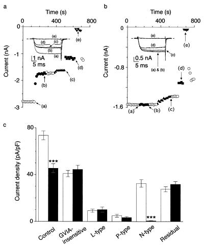
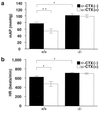
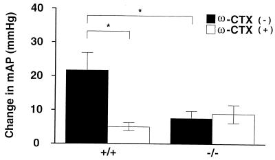
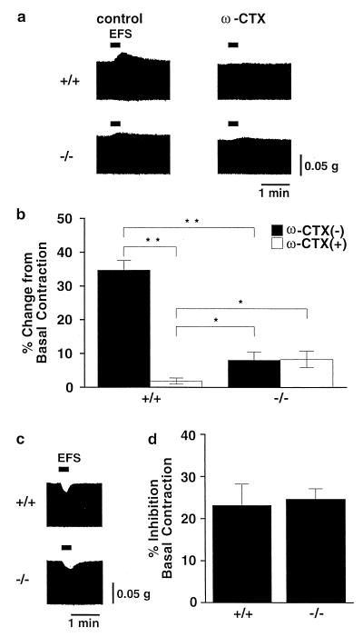
N-type voltage-dependent Ca(2+) channels (VDCCs), predominantly localized in the nervous system, have been considered to play an essential role in a variety of neuronal functions, including neurotransmitter release at sympathetic nerve terminals. As a direct approach to elucidating the physiological significance of N-type VDCCs, we have generated mice genetically deficient in the alpha(1B) subunit (Ca(v) 2.2). The alpha(1B)-deficient null mice, surprisingly, have a normal life span and are free from apparent behavioral defects. A complete and selective elimination of N-type currents, sensitive to omega-conotoxin GVIA, was observed without significant changes in the activity of other VDCC types in neuronal preparations of mutant mice. The baroreflex response, mediated by the sympathetic nervous system, was markedly reduced after bilateral carotid occlusion. In isolated left atria prepared from N-type-deficient mice, the positive inotropic responses to electrical sympathetic neuronal stimulation were dramatically decreased compared with those of normal mice. In contrast, parasympathetic nervous activity in the mutant mice was nearly identical to that of wild-type mice. Interestingly, the mutant mice showed sustained elevation of heart rate and blood pressure. These results provide direct evidence that N-type VDCCs are indispensable for the function of the sympathetic nervous system in circulatory regulation and indicate that N-type VDCC-deficient mice will be a useful model for studying disorders attributable to sympathetic nerve dysfunction.


Figures

Figure 1
Figure 1
Generation and characterization of mice lacking a functional α1B gene. (a) Strategy for gene disruption. B, BamHI site. (b) Southern blot analysis. Tail DNAs derived from heterozygous progeny were digested with BamHI. +/+, wild-type mice; 4.0 kb, KO mice (−/−); 3.1 kb, heterozygous mice (+/−). (c) Reverse transcription–PCR analysis of total brain RNA showed an alteration in fragment size. (Upper) Wild primer pair (P1 + P3). (Lower) Mutant primer pair (P2 + P3). m, 100-bp ladder. (d) Northern blot analysis of mouse poly(A)+ RNA demonstrates the absence of α1B mRNA in the brain of KO mice.
Figure 2
Figure 2
Histological analyses of brain in α1B-deficient mice. Cerebral cortex (a) and cerebellum (b) obtained from the mice (20 weeks) were dissected and stained with hematoxylin/eosin. [Bars: 50 μm (cerebral cortex) and 25 μm (cerebellum).]
Figure 3
Figure 3
Selective elimination of N-type channel current in SCG neurons of α1B-deficient mice. (a and b) Effects of 1 μM ω-CTX (●), 0.1 μM ω-agatoxin IVA (▴), 10 μM nimodipine (■), and 10 μM Cd2+ (▾) on Ba2+ currents in wild-type mice (a) and knock-out mice (b). (Insets) Representative current traces activated by depolarization for 30 ms to 5 mV from a Vh of −80 mV. (c) Histogram of current density in wild-type mice (open bar) and KO mice (solid bar). Data are expressed as means ± SE of 15 SCG neurons.
Figure 4
Figure 4
Response of mean arterial pressure and heart rate to (a and b) ω-CTX. Summarized results of mean arterial pressure (a) and heart rate (b) in the absence (solid column) or presence (open column) of ω-CTX (30 μg/kg) in wild-type and KO mice. mAP and HR were measured 30 min after administration of ω-CTX. Data are expressed as means ± SE.
Figure 5
Figure 5
Attenuation of vascular and cardiac sympathetic activities in N-type KO mice. Shown are the summarized results of blood pressure responses to bilateral carotid artery occlusion in the absence (solid column) or presence (open column) of ω-CTX in wild-type and KO mice. Each column represents the mean ± SE of five experiments.
Figure 6
Figure 6
Positive and negative inotropic responses to electrical field stimulation (EFS). (a) Representative records of positive inotropic responses to EFS in the absence (Left) or presence (Right) of ω-CTX (30 nM) in wild-type mice (Upper) and in N-type Ca2+ channel-deficient mice (Lower). Experiments were carried out in the presence of atropine (1 μM). (b) Summarized results of the sympathetic nerve-mediated positive inotropic responses in the presence or absence of ω-CTX. Each column represents the mean ± SE of five experiments. (c) Representative records of negative inotropic responses to EFS in wild-type mice (Upper) or KO mice (Lower). Experiments were carried out in the presence of propranolol (1 μM). (d) Summarized results of the parasympathetic nerve-mediated negative inotropic responses. Each column represents the mean ± SE of five experiments.

References

    1. Ertel E A, Campbell K P, Harpold M M, Hofmann F, Mori Y, Perez-Reyes E, Schwartz A, Snutch T P, Tanabe T, Birnbaumer L, et al. Neuron. 2000;25:533–535. - PubMed
    1. Olivera B M, Gray W R, Zeikus R, McIntosh J M, Varga J, River J, Santos V, Cruz L J. Science. 1985;230:1338–1343. - PubMed
    1. Dubel S J, Starr T V B, Hell J, Ahlijanian M K, Enyeart J J, Catterall W A, Snutch T P. Proc Natl Acad Sci USA. 1992;89:5058–5062. - PMC - PubMed
    1. Williams M W, Brust P F, Feldman D H, Patthi S, Simerson S, Maroufi A, Macue A F, Velicelebi G, Ellis S B, Harpold M M. Science. 1992;257:389–395. - PubMed
    1. Fujita Y, Mynlieff M, Dirksen R T, Kim M S, Niidome T, Nakai J, Friedrich T, Iwabe N, Miyata T, Furuichi T, et al. Neuron. 1993;10:585–598. - PubMed

MeSH terms