. 2001 Apr 24;98(9):5193-8.
doi: 10.1073/pnas.091484998. Epub 2001 Apr 10.

In vivo restoration of laminin 5 beta 3 expression and function in junctional epidermolysis bullosa

Affiliations

In vivo restoration of laminin 5 beta 3 expression and function in junctional epidermolysis bullosa

P B Robbins et al. Proc Natl Acad Sci U S A. .

Abstract

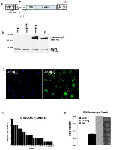
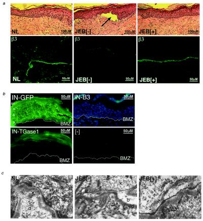
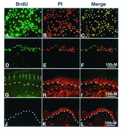
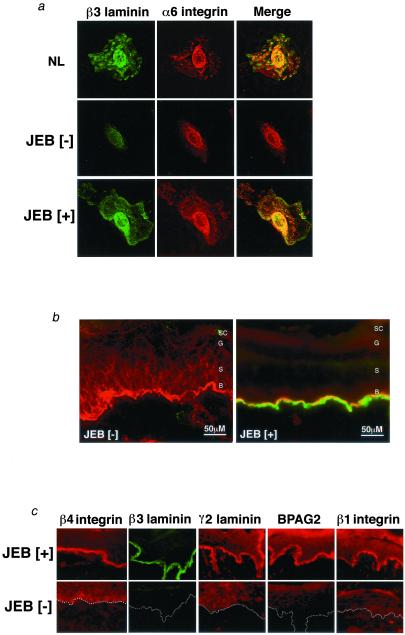
The blistering disorder, lethal junctional epidermolysis bullosa (JEB), can result from mutations in the LAMB3 gene, which encodes laminin 5 beta3 (beta3). Appropriate expression of LAMbeta3 in JEB skin tissue could potentially ameliorate the symptoms of the underlying disease. To explore the utility of this therapeutic approach, primary keratinocytes from six unrelated JEB patients were transduced with a retroviral vector encoding beta3 and used to regenerate human skin on severe combined immunodeficient (SCID) mice. Tissue regenerated from beta3-transduced JEB keratinocytes produced phenotypically normal skin characterized by sustained beta3 expression and the formation of hemidesmosomes. Additionally, beta3 gene transfer corrected the distribution of a number of important basement membrane zone proteins including BPAG2, integrins beta4/beta1, and laminins alpha3/gamma2. Skin produced from beta3-negative (beta3[-]) JEB cells mimicked the hallmarks of the disease state and did not exhibit any of the aforementioned traits. Therefore, by effecting therapeutic gene transfer to beta3-deficient primary keratinocytes, it is possible to produce healthy, normal skin tissue in vivo. These data support the utility of gene therapy for JEB and highlight the potential for gene delivery in the treatment of human genetic skin disease.


Figures

Figure 1
Figure 1
Laminin 5 β3 gene transfer. (a) Retroviral vector (IN-β3) generated for these studies. Expression of the human LAMB3 gene cDNA is driven via the internal cytomegalovirus (CMV) promoter. The IN-β3 vector has deletions of LTR sequences in the U3 region of the 3′ LTR previously implicated in retroviral silencing. (b) β3 protein expression following gene transfer to JEB patient cells. Primary JEB keratinocytes were transduced with either IN-β3 or GFP control vectors. Protein extracts from media were prepared 48 h later and subjected to Western blot analysis with the K140 monoclonal antibody to β3. MMP2 was included as an internal control of protein quality and loading. (c) Immunohistochemistry with β3 antibodies demonstrating efficiency of β3 gene transfer to JEB cells. Note the β3 expression (green) in all JEB cells after gene transfer (JEB[+]) compared with control (JEB[−]). Cell nuclei are counterstained with Hoechst 33342 (blue). (d) Distribution of the number proviral integrants in 24 transduced JEB cell holoclones. JEB cells were cloned at limiting dilution after IN-β3 transduction and 24 holoclones (35) were obtained. The presence and number of proviral genomes is noted for each clone. (e) Growth of JEB cells after β3 gene transfer (JEB[+]) or GFP gene transfer (JEB[−]) in comparison to normal keratinocytes (NL). Cells were plated at identical densities in triplicate and grown on tissue culture plastic for 4 days then counted.
Figure 2
Figure 2
Restoration of laminin 5 β3 to JEB skin in vivo. Genetically engineered human JEB skin was excised for examination 4 to 8 weeks after grafting and regeneration on SCID mice. (a) Histology of JEB skin in vivo (Upper)—note the normal histologic appearance with the presence of differentiated cell layers and the blister present in uncorrected skin (arrow in Center). NL, skin graft regenerated from normal keratinocytes; JEB[−], regenerated JEB tissue following GFP gene transfer; JEB[+], regenerated JEB tissue following β3 gene transfer. Restoration of correctly localized β3 protein expression in vivo (Lower). Data are representative of those obtained from tissue regenerated from six patients. (b) IN vector backbone drives gene expression throughout stratified epithelium. Epidermis was regenerated in vivo from JEB[−] keratinocytes transduced with either the IN-GFP or IN-β3 retrovector and fluorescence microscopy performed on unfixed cryosections. Note the GFP expression in all epidermal layers of IN-GFP[+] tissue and the lack of GFP expression in the IN-β3[+] tissue. Dual fluorescence (GFP + Hoechst 33342) staining of IN-β3 transduced tissue is shown to highlight cellular nuclei and the lack of GFP fluorescence. Epidermal layers are labeled: SC, stratum corneum; G, granular layer; S, spinous layer; B, basal layer. When using the same IN-vector backbone to drive expression of TGase-1 in regenerated LI patient skin, TGase-1 is only detectable in the stratum corneum (Lower Left) and absent in the nontransduced LI tissue (Lower Right). (c) Hemidesmosome formation in JEB skin after β3 gene transfer. Transmission electron microscopy was performed on skin regenerated from normal keratinocytes (NL), GFP transduced JEB tissue (JEB[−]), and β3-engineered JEB skin tissue (JEB[+]). Note the ultrastructural evidence of blister formation in uncorrected tissue and the lack of hemidesmosomes in JEB[−] tissue as opposed to the restoration of hemidesmosome structures (denoted by arrows) in JEB[+] tissue; d, dermis; ld, lamina densa; k, keratinocyte; b, blister. (×40,000 magnification.)
Figure 4
Figure 4
Kinetics of BrdUrd turnover in epidermal regeneration. Human keratinocytes were grown in the presence of BrdUrd for 5 days until all cells incorporated this nucleoside. Labeled cells were seeded on dermal substrate in vitro then grafted onto SCID mice in vivo. (AC) BrdUrd-labeled human keratinocytes in vitro; BrdUrd, immunostaining with FITC labeled antibody to BrdUrd; PI, propidium iodide counterstaining to highlight all of the cells present in the same field; Merge, merged image of identical fields subjected to BrdUrd and PI staining. (DF) Labeled human keratinocytes on dermal substrate in vitro. (GI) Skin regenerated from labeled cells at 3 weeks postgrafting. Note the layer of labeled cells in the outer epidermis in G (arrows). (JL) Skin regenerated from labeled cells at 6 weeks postgrafting. Note the loss of BrdUrd detection by 6 weeks postgrafting, which corresponds to 8 weeks postlabeling. The dashed line in GL denotes the basement membrane zone.
Figure 3
Figure 3
Laminin β3 delivery restores polarized distribution of protein components of the epidermal basement membrane. (a) Expression of laminin 5 β3 (green) and α6 integrin (red) was examined in β3-transduced (JEB[+]) and untransduced JEB cells (JEB[−]), as well as normal control (NL) cells by laser confocal immunofluorescence microscopy. Note the diminished presence of α6 integrin with a perinuclear distribution in the JEB[−] cell and the restoration of appropriately distributed expression concomitant with restoration of β3 expression. (b) Expression of laminin 5 β3 (green) and α6 integrin (red) was examined in β3-engineered JEB skin tissue (JEB[+]) and GFP-transduced control (JEB[−]) by laser confocal microscopy. Note the mislocalization of α6 integrin throughout the epidermis in the absence of β3 in JEB[−] tissue and the restoration of strongly polarized distribution to the BMZ with β3 restoration. (c) Localization and expression of basement membrane proteins integrins β4 and α6, laminins β1, β3, and γ2, and BPAG2 were examined as a function of laminin 5 β3 delivery in β3-engineered JEB skin tissue (JEB[+]) and GFP-transduced control (JEB[−]) tissue. Note the decreased and/or mislocalized distribution of all of the BMZ proteins in JEB[−] tissue and the restoration of polarized expression after β3 gene delivery. Data are representative of independently analyzed tissues generated from cells of multiple patients. Dotted lines denote the location of the BMZ in JEB[−] tissue.

References

    1. Uitto J, Eady R, Fine J D, Feder M, Dart J. J Invest Dermatol. 2000;114:734–737. - PubMed
    1. Akiyama M. Int J Dermatol. 1998;37:722–728. - PubMed
    1. Bale S J, Doyle S Z. J Invest Dermatol. 1994;102:49S–50S. - PubMed
    1. Uitto J, Pulkkinen L, McLean W H. Mol Med Today. 1997;3:457–465. - PubMed
    1. Marinkovich M P. Dermatol Clin. 1999;17:473–485. - PubMed

Publication types

MeSH terms

Substances