. 2001 Apr 10;98(8):4528-33.
doi: 10.1073/pnas.071002398.

Spatial and temporal emergence of high proliferative potential hematopoietic precursors during murine embryogenesis

Affiliations

Spatial and temporal emergence of high proliferative potential hematopoietic precursors during murine embryogenesis

J Palis et al. Proc Natl Acad Sci U S A. .

Abstract

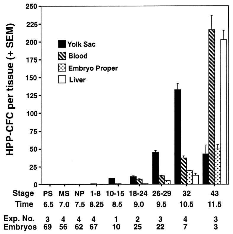
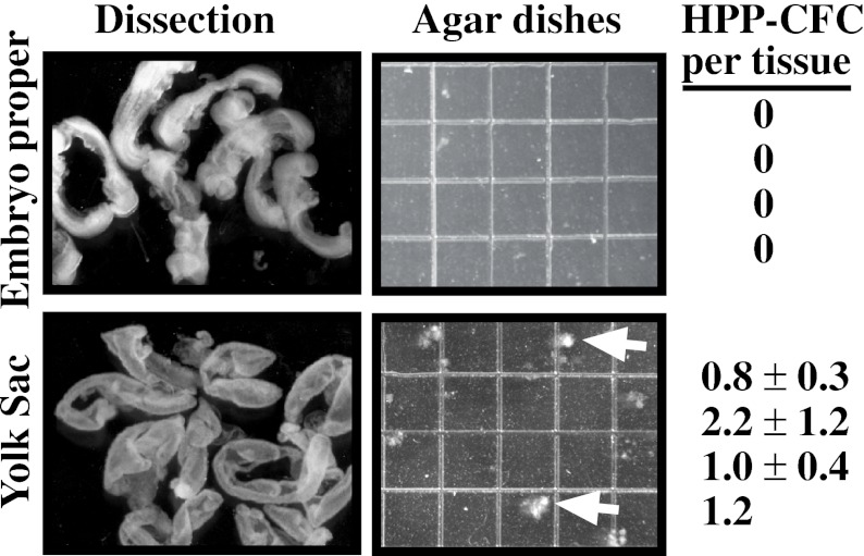
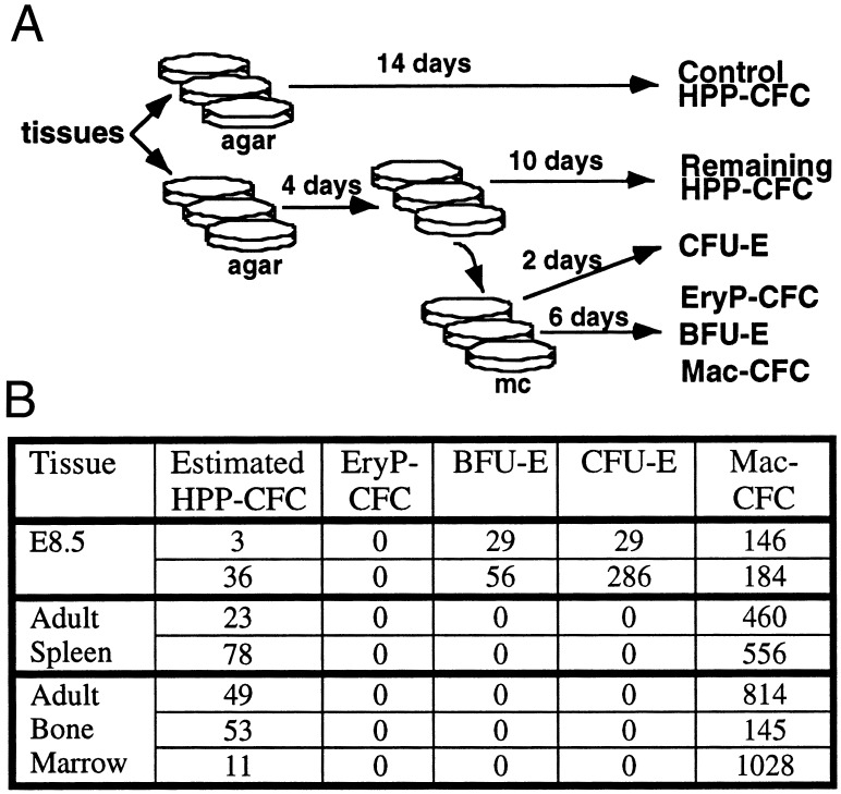
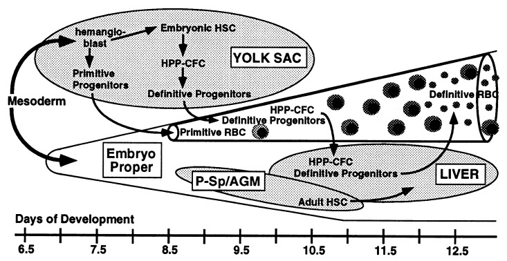
During mouse embryogenesis, two waves of hematopoietic progenitors originate in the yolk sac. The first wave consists of primitive erythroid progenitors that arise at embryonic day 7.0 (E7.0), whereas the second wave consists of definitive erythroid progenitors that arise at E8.25. To determine whether these unilineage hematopoietic progenitors arise from multipotential precursors, we investigated the kinetics of high proliferative potential colony-forming cells (HPP-CFC), multipotent precursors that give rise to macroscopic colonies when cultured in vitro. No HPP-CFC were found at presomite stages (E6.5-E7.5). Rather, HPP-CFC were detected first at early somite stages (E8.25), exclusively in the yolk sac. HPP-CFC were found subsequently in the bloodstream at higher levels than the remainder of the embryo proper. However, the yolk sac remains the predominant site of HPP-CFC expansion (>100-fold) until the liver begins to serve as the major hematopoietic organ at E11.5. On secondary replating, embryonic HPP-CFC give rise to definitive erythroid and macrophage (but not primitive erythroid) progenitors. Our findings support the hypothesis that definitive but not primitive hematopoietic progenitors originate from yolk sac-derived HPP-CFC during late gastrulation.


Figures

Figure 1
Figure 1
Distribution of HPP-CFC (mean + SEM) within various compartments of staged mouse conceptuses during gastrulation and early organogenesis (E6.5–E11.5). The liver was assayed separately from the remainder of the embryo proper at 32 and 43 somite pairs. Representative results at each stage are shown; multiple independent experiments (Exp. No.) gave similar results. The total number of embryos (Embryos) investigated at each stage is also listed. PS, prestreak; MS, midprimitive streak; NP, neural plate.
Figure 2
Figure 2
Dissection of early somite-pair stage embryos (E8.25) into yolk sac and embryo proper tissues, and HPP-CFC (mean ± SEM) derived from those tissues. Grids on the agar dishes represent 2 mm. HPP-CFC are shown by white arrows. The number of HPP-CFC (mean ± SEM) per embryo proper and per yolk sac are shown for four independent experiments. HPP-CFC were plated in triplicate in three of four experiments.
Figure 3
Figure 3
(A) Experimental design for replating of day 4 HPP-CFC. Single-cell suspensions of various tissues were plated into six double agar dishes. At day 4, colonies >10 cells were plucked from three dishes, pooled, and replated into methylcellulose (mc) with growth factors that support both primitive erythroid (EryP-CFC), definitive erythroid (BFU-E and CFU-E), and macrophage (Mac-CFC) progenitor differentiation. HPP-CFC and committed hematopoietic progenitors were enumerated at the times shown. (B) Hematopoietic progenitors derived from day-4 colonies >10 cells (HPP-CFC) replated from early somite-pair embryos, adult spleen, and adult bone marrow. The number of replated HPP-CFC was estimated by the difference in HPP-CFC between control and remaining HPP-CFC dishes.
Figure 4
Figure 4
The temporal appearance of primitive erythroid progenitors and HPP-CFC derived from EBs differentiated for 3 to 10 days. Although the results of a representative experiment are shown, two other independent experiments gave similar results.
Figure 5
Figure 5
Model of early hematopoietic ontogeny in the mouse embryo. P-Sp/AGM, para-aortic splanchnopleura/aorta-gonad-mesonephros region; RBC, red blood cells.

Similar articles

Cited by

References

    1. Osawa M, Hanada K, Hamada H, Nakauchi H. Science. 1996;273:242–245. - PubMed
    1. Moore M A S. Blood. 1991;78:1–19. - PubMed
    1. Bertoncello I. Curr Top Microbiol Immunol. 1992;177:83–94. - PubMed
    1. Bradley T R, Hodgson G S. Blood. 1979;54:1446–1450. - PubMed
    1. McNiece I K, Williams N T, Johnson G R, Kriegler A B, Bradley T R, Hodgson G S. Exp Hematol (Charlottesville, Va) 1987;15:972–977. - PubMed

Publication types