. 2001 Jun 22;276(25):22341-50.
doi: 10.1074/jbc.M010839200. Epub 2001 Apr 10.

Transforming growth factor-beta - and tumor necrosis factor-alpha -mediated induction and proteolytic activation of MMP-9 in human skin

Affiliations

Transforming growth factor-beta - and tumor necrosis factor-alpha -mediated induction and proteolytic activation of MMP-9 in human skin

Y P Han et al. J Biol Chem. .

Abstract

Both cytokines and matrix metalloproteinases (MMPs) are active during physiologic and pathologic processes such as cancer metastasis and wound repair. We have systematically studied cytokine-mediated MMP regulation. Cytokine-mediated proteinase induction and activation were initially investigated in organ-cultured human skin followed by determination of underlying cellular and molecular mechanisms using isolated skin cells. In this report we demonstrate that tumor necrosis factor-alpha (TNF-alpha) and transforming growth factor-beta (TGF-beta) synergistically induce pro-MMP-9 in human skin as well as isolated dermal fibroblasts and epidermal keratinocytes. Furthermore, TNF-alpha promotes proteolytic activation of pro-MMP-9 by conversion of the 92-kDa pro-MMP-9 to the 82-kDa active enzyme. This activation occurred only in skin organ culture and not by either isolated fibroblasts or keratinocyte, although the pro-MMP-9 activation could be measured in a cell-free system derived from TNF-alpha-activated skin. The cytokine-mediated induction of pro-MMP-9 in dermal fibroblasts was evident by increased mRNA. At the transcription level, we examined the cytokine-mediated transactivation of the 5'-region promoter of the human MMP-9 in dermal fibroblasts. The results demonstrated that TNF-alpha and TGF-beta could independently stimulate the 5'-flanking 670-base pair promoter. A TGF-beta-response element (-474) and an NF-kappaB-binding site (-601) were identified to be the cis-elements for TGF-beta or TNF-alpha activation, respectively. Taken together, these findings suggest a specific mechanism whereby multiple cytokines can regulate MMP-9 expression/activation in the cells of human skin. These results imply roles for these cytokines in the regulation of MMP-9 in physiologic and pathologic tissue remodeling.


Figures

FIG. 1
FIG. 1. Cytokine-mediated induction and activation of MMP-9 in organ-cultured human skin
Normal full thickness human skin was cultured in serum-free DMEM and stimulated by TNF-α (10 ng/ml) and TGF-β (1 ng/ml) either individually or combined. Skin samples from eight distinct individuals were examined for cytokine-mediated induction and activation of MMP-9. All showed similar results. A, conditioned media were sampled at the indicated time points and analyzed for gelatinolytic activities by zymogram. The 92- and 82-kDa gelatinolytic activities are indicated by arrows. B, identities of the 92- and 82-kDa gelatinolytic activities as pro-MMP-9 and active MMP-9 were determined by Western blot. The total gelatinase from 500-µl conditioned media derived from 72-h culture was enriched by gelatin-conjugated Sepharose 4B matrix. The bound protein was resolved by SDS-PAGE and detected by Western blot using polyclonal anti-MMP-9 antibodies.
FIG. 2
FIG. 2. TNF-α-mediated pro-MMP-9 activation occurs in the skin tissue but not in the medium
Human skin was cultured in DMEM with or without TNF-α. After 70 h of incubation the conditioned media were collected. The skin tissues were washed and replanted in the fresh DMEM. The original conditioned media and the replanted skin tissue in DMEM were incubated at 37 °C, and media were sampled at the indicated time points. MMP-9 activities were measured by gelatinolytic zymogram.
FIG. 3
FIG. 3. Proteolytic activation of pro-MMP-9 in the cell-free skin extract
Adult human skin was stimulated with TNF-α (10 ng/ml) in DMEM for 70 h. The tissue was washed and minced following extraction using 2% Triton X-100 (see “Experimental Procedures”). The detergent-soluble and -insoluble fractions were separated by centrifugation. Purified pro-MMP-9 was added or not to these fractions and incubated at 37 °C for 20 h. MMP-9 activities were measured by gelatinolytic zymogram.
FIG. 4
FIG. 4. Cytokine-mediated induction of pro-MMP-9 in human dermal fibroblasts and epidermal keratinocyte
A, dermal fibroblasts were isolated from the adult human skin, and early passages were embedded into type I collagen. The cells were cultured in serum-free DMEM and stimulated with TNF-α, TGF-β, PDGF-BB, EGF, IL-8, and IL-6 at 10 ng/ml. In another panel the cells were stimulated with a combination of TGF-β with TNF-α, PDGF, EFG, IL-8, or IL-6, all at 10 ng/ml. After cultivation for 68 h the conditioned media were analyzed for gelatinolytic zymography. The 92-kDa MMP-9 and 72- and 62-kDa MMP-2 are indicated. B, the identity of the 92-kDa gelatinase activity was confirmed as MMP-9. The total gelatinase from 500-µl conditioned media was enriched by gelatin-conjugated Sepharose 4B matrix. The bound protein was resolved by SDS-PAGE and detected for MMP-9 by Western blot. C, the keratinocytes were isolated from the epidermal portion of adult human skin and the early passages were used for the experiment. The confluent monolayers were stimulated by TNF-α and TGF-β individually or combined in basal medium (KBM) for 70 h. A typical zymography is presented.
FIG. 5
FIG. 5. Time course of co-induction of pro-MMP-9 by TNF-α and TGF-β
The early passages of human dermal fibroblasts were cultured as monolayers or embedded in type I collagen. The cells were stimulated by a combination of TNF-α (10 ng/ml) and TGF-β (10 ng/ml) in DMEM. A, the fibroblast monolayers were stimulated by the combined cytokines, and conditioned media was sampled at the indicated time points and analyzed for gelatinolytic activities. B, the fibroblasts embedded in collagen lattices were stimulated by the cytokines, and conditioned media were assayed for gelatinolytic activities.
FIG. 6
FIG. 6. Concentration dependence of the cytokine-mediated induction of pro-MMP-9
The human dermal fibroblasts were cultured as monolayers. One set of six wells was stimulated with TNF-α at 10 ng/ml and with varying concentrations of TGF-β as indicated. Another set of six wells was stimulated by TGF-β at 10 ng/ml and varying concentrations of TNF-α. After stimulation for 48 h the conditioned media were analyzed for gelatinolytic zymography.
FIG. 7
FIG. 7. The cytokine-mediated induction of MMP-9 is up-regulated at the mRNA level
A, the human dermal fibroblasts as monolayers were stimulated by TNF-α (10 ng/ml) and TGF-β (10 ng/ml) individually or combined in serum-free DMEM. After stimulation for 20 h the total RNA was extracted. The mRNA levels of MMP-9 and β-actin were determined by RT-PCR. For the β-actin, one-third of PCR product was loaded into the gel as shown. The identity of PCR product for MMP-9 was confirmed by its expected size and DNA sequencing. B, time course of MMP-9 mRNA co-induction by the two cytokines. The mRNA level of MMP-9 and β-actin was measured at the indicated time points, and the relative amount was quantitated by Alpha-imaging.
FIG. 8
FIG. 8. Actinomycin D and cycloheximide attenuates the cytokine-mediated induction of pro-MMP-9 in dermal fibroblasts
The dermal fibroblasts were embedded in collagen and stimulated with or without TNF-α (10 ng/ml) and TGF-β (1 ng/ml) as indicated. Actino-mycin D (Act D) and cycloheximide (CHX) were applied at the indicated concentration in the DMEM. After 70-h culture the conditioned media were analyzed for gelatinolytic zymography.
FIG. 9
FIG. 9. TNF-α and TGF-β activates the human MMP-9 promoter
The human dermal fibroblasts were transiently transfected by pMMP9–670-CAT, which contains 670 bp of 5′-promoter of the human MMP-9 gene. After a 3-h transfection the cells were stimulated by TNF-α (10 ng/ml) and TGF-β (1 ng/ml) as indicated for an additional 24, 48, and 68 h. The promoter activation was measured by CAT assay. A, the CAT activities expressed in the cells are shown as the percentage of acetylated products measured by PhosphorImaging and are the average of three replicates. B, the conditioned medium of the transient transfected cells was analyzed for gelatinolytic activities, which represent the endogenous MMP-9 induction.
FIG. 10
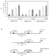
FIG. 10. One of the two potential NF-κB-responsive elements in the MMP-9 promoter is essential for TNF-α -mediated promoter activation
The dermal fibroblasts were transiently transfected with pM9–670-CAT (wild type), pM9–590-CAT (p65NF-κB delete), and pM9–670-mp50-CAT (p50NF-κB site mutant). After transfection for 3 h the cells were stimulated with the cytokines individually or combined. A, the CAT activities expressed in the cells were measured. The values represent the mean of results obtained from three replicate culture wells. B, illustration of the construction used in the experiments. The potential p65 and p50 NF-κB-binding sites, and the mutant version are illustrated.
FIG. 11
FIG. 11. TGF-β activates the minimal promoter of the human MMP-9 through the TRE-response site
The human dermal fibroblasts were transiently transfected by the CAT reporter plasmids, pM9–670-CAT (wild type), pM9–670-mTRE-CAT (TRE site mutant), and pM9–460-CAT (p65 NF-κB and TRE delete). The cells were stimulated by cytokines individually or combined. A, the CAT activities from the cells were measured. The mean value of triplicate experiments is indicated for each reporter plasmid. B, the plasmid constructs and the potential cytokine-response elements are illustrated.

Similar articles

Cited by

References

    1. Leibovich SJ, Polverini PJ, Shepard HM, Wiseman DM, Shively V, Nuseir N. Nature. 1987;329:630–632. - PubMed
    1. Fajardo LF, Kwan HH, Kowalski J, Prionas SD, Allison AC. Am. J. Pathol. 1992;140:539–544. - PMC - PubMed
    1. Garner WL, Karmiol S, Rodriguez JL, Smith DJ, Jr, Phan SH. J. Invest. Dermatol. 1993;101:875–879. - PubMed
    1. Cooney R, Iocono J, Maish G, Smith JS, Ehrlich P. J. Trauma. 1997;42:415–420. - PubMed
    1. Kitzis V, Engrav LH, Quinn LS. J. Surg. Res. 1999;87:134–141. - PubMed

Publication types

MeSH terms

LinkOut - more resources