. 2001 Apr;31(4):1261-7.
doi: 10.1002/1521-4141(200104)31:4<1261::AID-IMMU1261gt;3.0.CO;2-H.

Ontogeny, distribution and function of CD38-expressing B lymphocytes in mice

Affiliations
Free PMC article

Ontogeny, distribution and function of CD38-expressing B lymphocytes in mice

F R Donís-Hernández et al. Eur J Immunol. 2001 Apr.
Free PMC article

Abstract

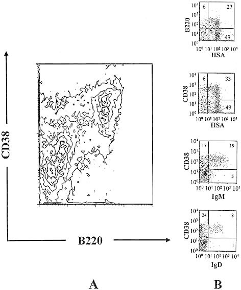
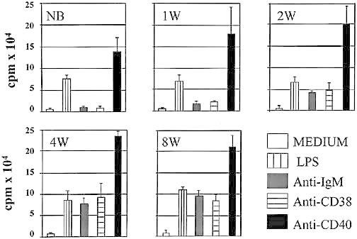
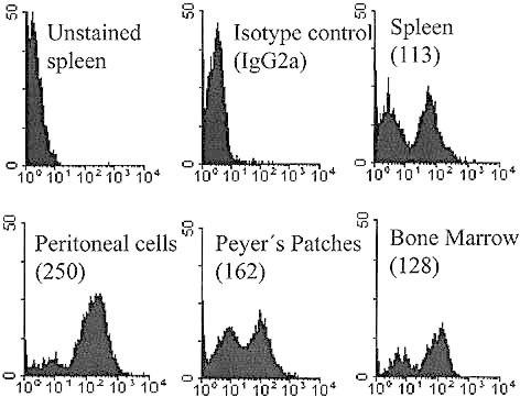
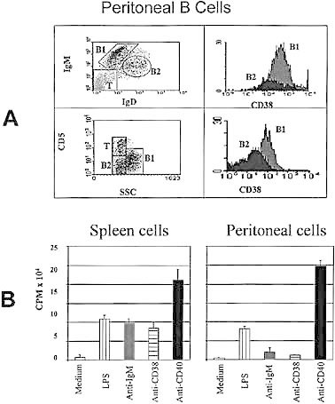
Analysis of expression of CD38, CD45R (B220), IgM and IgD on splenic B lymphocytes from mice of different ages demonstrated CD38 on both immature (B220(+), BCR(-)) and mature (B220(+), BCR(+)) B lymphocytes. Similarly, CD38 is expressed as early as B220 on the surface of progenitor B cells in the bone marrow. In spite of expressing of CD38 and IgM, neonatal B cells, in contrast to the adult, failed to proliferate to either anti-CD38 or anti-IgM cross-linking when IL-4 was present. They did, however, respond to LPS and anti-CD40, and by 2 weeks of age they began to respond to anti-CD38 and anti-IgM, reaching adult B cell levels by 4 weeks. Although the distribution of CD38 on adult B cells from most different lymphoid compartments was broadly similar, significantly higher levels of CD38 were expressed on peritoneal B lymphocytes. A detailed analysis, using IgM / IgD ratio and staining with anti-CD5 confirmed that B1 lymphocytes were expressing a high level of CD38. Interestingly, both immature B cells and peritoneal B1 lymphocytes were unresponsive to anti-CD38. However, they were activated by LPS or anti-CD40.


Figures

Fig. 1
Fig. 1
Ontogeny of the expression of CD38 in the spleen. Splenic lymphocytes from BALB / c mice at different ages were stained with B220-FITC (A), anti-IgM-FITC (B) or anti-IgD-FITC (C) and counter-stained with anti-CD38-PE. IC: isotype control staining for the pairs used in each combination; NB: newborn mice; 1 – 8W, 1 – 8 weeks old.
Fig. 2
Fig. 2
Expression of CD38 in bone marrow from adult mice. Bone marrow cells from 8-week-old BALB / c mice were stained with B220-FITC and anti-CD38-PE for panel A; and anti-HSA-FITC, anti-IgM-FITC or anti-IgD-FITC and then counter-stained with B220-PE or anti-CD38-PE for panel B.
Fig. 3
Fig. 3
Immature B lymphocytes fail to proliferate to anti-CD38 stimulation. 106-MACS purified splenic B lymphocytes from BALB / c mice at different ages were stimulated with 50 μg / ml LPS, 10 μg / ml anti-mouse IgM (B7.6), 20 μg / ml anti-mouse CD38 (NIM-R5), or 10 μg / ml anti-mouse CD40 (1C10). All cultures contained 10 U / ml of recombinant mouse IL-4. The cultures were incubated for 72 h at 37 °C, 5 % CO2 and pulsed with [3H] thymidine during the last 4 h of culture. Results are shown as mean cpm plus standard deviation from four wells. This is a representative experiment of five done in the same manner and with similar results.
Fig. 4
Fig. 4
B lymphocytes from three strains of adult mice express and proliferate to anti-CD38 stimulation. Splenic lymphocytes from three strains of adult mice were stained with anti-B220-FITC / anti-CD38-PE (panel A). Purified B lymphocytes from spleen of each strain of mice were stimulated with 10 μg / ml anti-mouse IgM (B7.6), 20 μg / ml anti-mouse CD38 (NIM-R5), or 10 μg / ml anti-mouse CD40 (1C10). All cultures contained 10 U / ml of recombinant mouse IL-4. The cultures were incubated for 72 h at 37 °C, 5 % CO2 and pulsed with [3H] thymidine during the last 4 h of culture. Results are shown as mean cpm plus standard deviation from 4 wells (panel B). This is a representative experiments of five done in the same manner and with similar results.
Fig. 5
Fig. 5
Differential expression of CD38 on B lymphocytes from different lymphoid compartments. Total lymphocytes from spleen, peritoneal cavity, Peyer's patches and bone marrow were stained with FITC-anti-mouse CD38 (NIM-R5). Numbers in parentheses show the mean fluorescence intensity of the CD38+ cells.
Fig. 6
Fig. 6
B1 lymphocytes from peritoneal cavity express high levels of CD38 on their surface, but fail to proliferate to CD38 cross-linking. Lymphocytes from the peritoneal cavity were stained with anti-IgD-FITC / anti-IgM-PE, anti-CD38-biotin and then streptavidin-PerCP or with anti-CD38-FITC / anti-CD5-PE (panel A). Purified B lyphocytes from the peritoneal cavity or the spleen were stimulated with 50 μg / ml LPS, 10 μg / ml anti-mouse IgM (B7.6), 20 μg / ml anti-mouse CD38 (NIM-R5), or 10 μg / ml anti-mouse CD40 (1C10). All cultures contained 10 U / ml of recombinant mouse IL-4. The cultures were incubated for 72 h at 37 °C, 5 % CO2 and pulsed with [3H] thymidine during the last 4 h of culture. Results are shown are mean cpm plus standard deviation from 4 wells (panel B). This is a representative experiments of five done in the same manner and with similar results.

References

    1. Lund FE, Cockayne DA, Randall TD, Solvason N, Schuber F, Howard MC. CD38: a new paradigm in lymphocyte activation and signal transduction. Immunol. Rev. 1998;161:79–93. - PubMed
    1. Ferrero E, Saccucci F, Malavasi F. The making of a leukocyte receptor: origin, genes and regulation of human CD38 and related molecules. Chem. Immunol. 2000;75:1–19. - PubMed
    1. Campana D, Suzuki T, Todisco E, Kitanaka A. CD38 in hematopoiesis. Chem. Immunol. 2000;75:169–188. - PubMed
    1. Konopleva M, Rissling I, Andreeff M. CD38 in hematopoietic malignancies. Chem. Immunol. 2000;75:189–206. - PubMed
    1. Bofill M, Borthwick NJ. CD38 in health and disease. Chem. Immunol. 2000;75:218–234. - PubMed

Publication types

MeSH terms