. 2001 Apr;13(7):1420-8.
doi: 10.1046/j.0953-816x.2001.01517.x.

NMDA-evoked calcium transients and currents in the suprachiasmatic nucleus: gating by the circadian system

Affiliations

NMDA-evoked calcium transients and currents in the suprachiasmatic nucleus: gating by the circadian system

C S Colwell. Eur J Neurosci. 2001 Apr.

Abstract

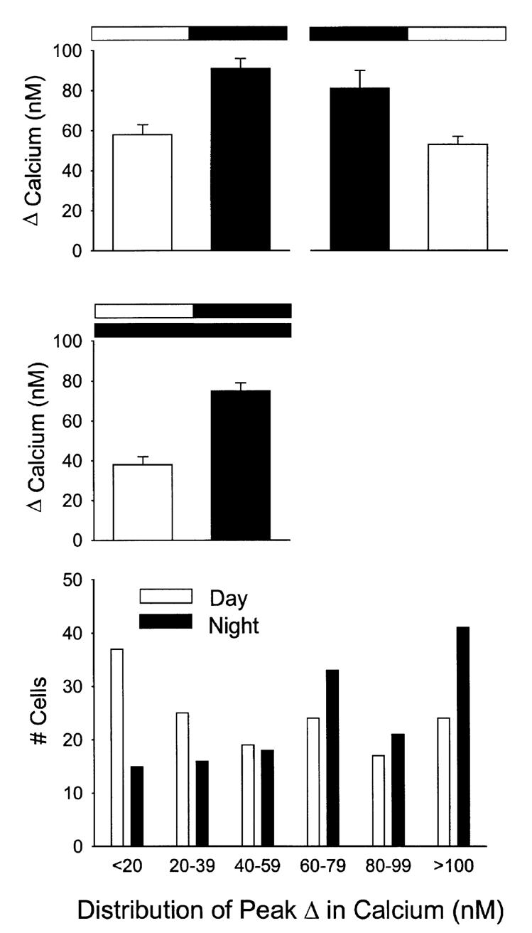
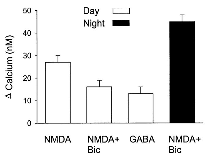
A variety of evidence suggests that the effects of light on the mammalian circadian system are mediated by glutamatergic mechanisms and that the N-methyl- D-aspartate (NMDA) receptor plays an important role in this regulation. One of the fundamental features of circadian oscillators is that their response to environmental stimulation varies depending on the phase of the daily cycle when the stimuli are applied. For example, the same light treatment, which can produce phase shifts of the oscillator when applied during subjective night, has no effect when applied during the subjective day in animals held in constant darkness (DD). We examined the hypothesis that the effects of NMDA on neurons in the suprachiasmatic nucleus (SCN) also vary from day to night. Optical techniques were utilized to estimate NMDA-induced calcium (Ca2+) changes in SCN cells. The resulting data indicate that there was a daily rhythm in the magnitude and duration of NMDA-induced Ca2+ transients. The phase of this rhythm was determined by the light-dark cycle to which the rats were exposed with the Ca2+ transients peaking during the night. This rhythm continued when animals were held in DD. gamma-Aminobutyric acid (GABA)ergic mechanisms modulated the NMDA response but were not responsible for the rhythm. Finally, there was a rhythm in NMDA-evoked currents in SCN neurons that also peaked during the night. This study provides the first evidence for a circadian oscillation in NMDA-evoked Ca2+ transients in SCN cells. This rhythm may play an important role in determining the periodic sensitivity of the circadian systems response to light.


Figures

FIG. 1
FIG. 1
Examples of Ca2+ transients measured from SCN cells in a brain slice loaded with the Ca2+ indicator dye fura2. Each line represents data collected from an individual cell. All of this data was collected in the absence of Mg2+ but in the presence of TTX and methoxyverapamil that were included to block voltage-sensitive Na+ and Ca2+ currents. Top: SCN cells show an increase in Ca2+ in response to bath application of NMDA (50 μM, 40 s). Bottom: NMDA-induced Ca2+ transients were inhibited by the presence of the NMDA receptor antagonist AP5 (50 μM). Data collected during the night from single SCN slice from a 14-day-old rat.
FIG. 2
FIG. 2
NMDA regulation of Ca2+ levels in the SCN. Bath application of NMDA (50 μM, 60 s) produced Ca2+ transients in most cells (94%) within the SCN (P < 0.001, n = 64). These NMDA-induced Ca2+ transients were blocked by treatment with the NMDA GluR antagonist AP5 (50 μM, P < 0.001, n = 32) and were enhanced by the removal of magnesium (Mg2+) from the extracellular solution (P < 0.001, n = 39). Finally, because Ca2+ influx through voltage-activated channels might be a major source of NMDA-evoked Ca2+ increases, some experiments were run in the presence of the TTX and methoxyverapamil (D600). In the presence of these channel blockers and the absence of Mg2+, bath application of NMDA (50 μM) still produced significant Ca2+ transients (P < 0.001, n = 83). Data collected during day from 10–15-day-old rats.
FIG. 3
FIG. 3
Diurnal rhythm in NMDA-evoked Ca2+ transients in SCN cells. In these experiments, NMDA-evoked Ca2+ transients were measured in SCN neurons in brain slices from animals during their day and compared to data obtained from brain slices from animals during their night. Animals were killed at either ZT 0 for the day group or ZT 12 for the night group. Each cell was sampled only once. Top left panel: NMDA-evoked Ca2+ transients peaked during night (day n = 83, night n = 102, P < 0.001). Top right panel: when the phase of the light-dark cycle to which the animals were exposed was reversed, so did the resulting rhythm (day n = 72; night n = 65, P < 0.05). Middle panel: in these experiments, animals were maintained in constant conditions and NMDA-evoked Ca2+ transients were measured in SCN neurons in brain slices from animals during their subjective day and compared to data obtained from brain slices from animals during their subjective night. Once again, NMDA-evoked Ca2+ transients peaked during subjective night (day n = 64; night n = 89, P < 0.001). Bottom panel: histogram illustrates the daily variation in the distribution of the NMDA-induced Ca2+responses. As has been previously reported (Colwell, 2000), in the presence of the ion channel blockers (TTX, D600), there was no significant day/night variation in the resting Ca2+ levels (day 89 ± 2 nM, n = 83; night 83 ± 1 nM, n = 102).
FIG. 4
FIG. 4
GABAnergic modulation of NMDA-evoked Ca2+ transients in cells in the SCN. During the day, in the presence of TTX and methoxyverapamil, NMDA-evoked Ca2+ transients were reduced by the application of the GABAA antagonist bicuculline (bicuculline + NMDA n = 39; NMDA n = 32, P < 0.05). This effect of bicuculline (20 μM) was not seen when experiments were run in the absence of extracellular Mg2+. By itself, bath application of GABA (100 μM) produced a modest increase in Ca2+ (n = 197). This effect of GABA was extremely variable as 44% of the cells sampled showed a Ca2+ increase (> 5%) in response to GABA whereas 21% showed a decrease in Ca2+. In an experiment run in the presence of bicuculline, it was still possible to see a day—night difference in the magnitude of NMDA-induced Ca2+ transients (day n = 39; night n = 46; P < 0.01).
FIG. 5
FIG. 5
Daily rhythm in NMDA currents in SCN neurons. Whole-cell patch clamp recording techniques were used to directly measure currents evoked by NMDA in SCN neurons. The voltage-dependence of the NMDA-evoked currents was measured by moving the cell through a ramp of voltages (schematically illustrated in the top insert) before, during, and after treatment with NMDA in the bath. As with the imaging experiments, these responses were measured in the presence of TTX and methoxyverapamil. Top panel shows an NMDA response recorded using this protocol. All data were collected as the cell’s membrane potential was moved from +40 mV to –90 mV. Bottom panel: NMDA currents were recorded in neurons in brain slices taken from animals during their day and compared to data obtained from brain slices from animals during their night. These experiments were run in the absence of Mg2+ to reduce the voltage-dependence of the NMDA response and in the presence of cadmium (Cd2+, 25 μM), and TTX (1 μM) in the bath. Under these conditions, there was a daily rhythm in NMDA-evoked inward currents (INMDA + IK) with peak responses significantly (P < 0.001) higher during the night (n = 14) than during the day (n = 11). In other experiments, voltage-gated K+ currents were also blocked with TEA in the bath and Cs+ (125 mM) in the patch pipette. Under these conditions, there was also a daily rhythm in NMDA-evoked inward currents (INMDA) with peak responses significantly (P < 0.05) higher during the night (n = 21) than during the day (n = 23).

References

    1. Alberi S, Dauphin MD, Dreifuss JJ, Raggenbass M. Whole-cell NMDA-evoked current in suprachiasmatic neurones of the rat: modulation by extracellular calcium ions. Brain Res. 1997;745:55–66. - PubMed
    1. Arendt J. Biological rhythms: the science of chronobiology. J. Royal College Physicians London. 1998;32:27–35. - PMC - PubMed
    1. Block GD, Khalsa SB, McMahon DG, Michel S, Guesz M. Biological clocks in the retina: cellular mechanisms of biological timekeeping. Int. Rev. Cytology. 1993;146:83–144. - PubMed
    1. Burgard EC, Hablitz JJ. Developmental changes in the voltage-dependence of neocortical NMDA responses. Dev. Brain Res. 1994;80:275–278. - PubMed
    1. Cagampang FR, Rattray M, Campbell IC, Powell JF, Coen CW. Variation in the expression of the mRNA for protein kinase C isoforms in the rat suprachiasmatic nuclei, caudate putamen and cerebral cortex. Mol. Brain Res. 1998;53:277–284. - PubMed

Publication types

MeSH terms