. 2001;3(3):168-77.
doi: 10.1186/ar294. Epub 2001 Jan 26.

IL-17 derived from juxta-articular bone and synovium contributes to joint degradation in rheumatoid arthritis

Affiliations

IL-17 derived from juxta-articular bone and synovium contributes to joint degradation in rheumatoid arthritis

M Chabaud et al. Arthritis Res. 2001.

Abstract

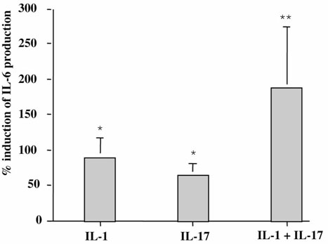
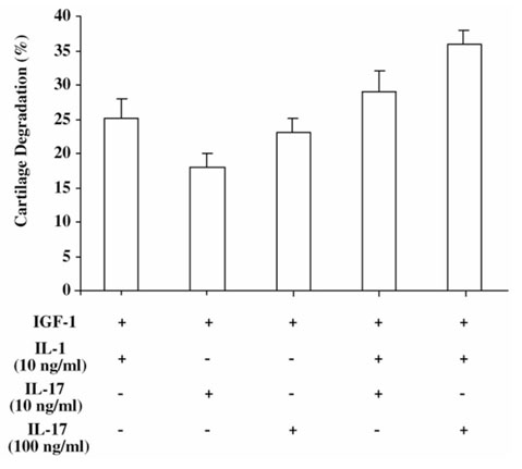
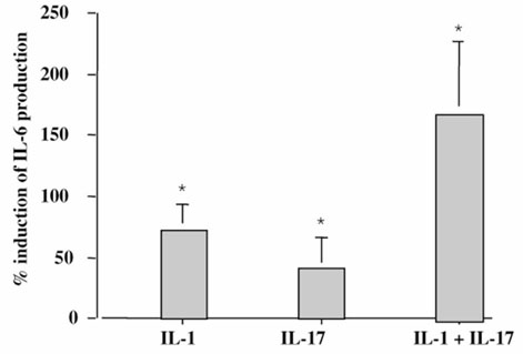
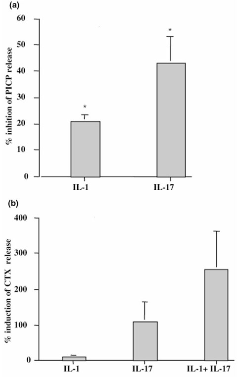
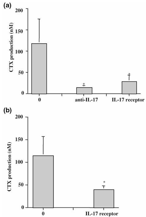
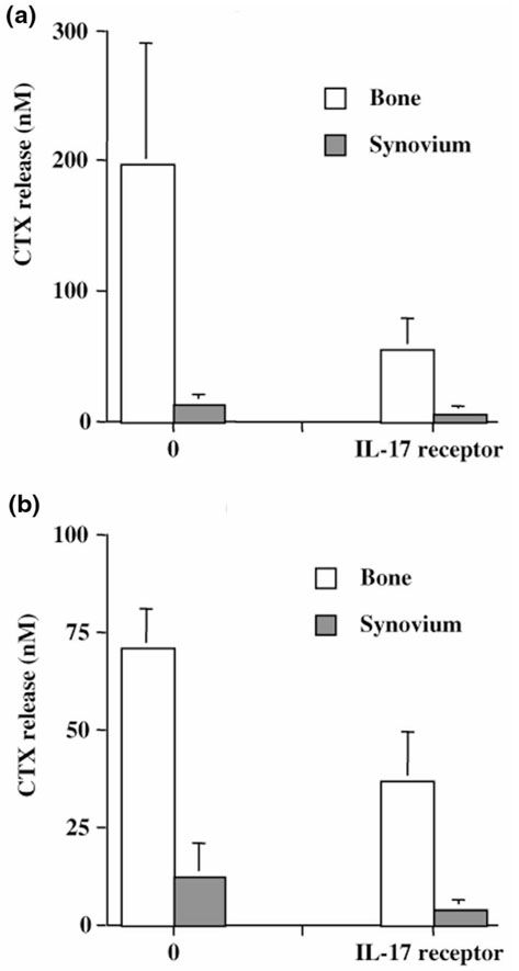
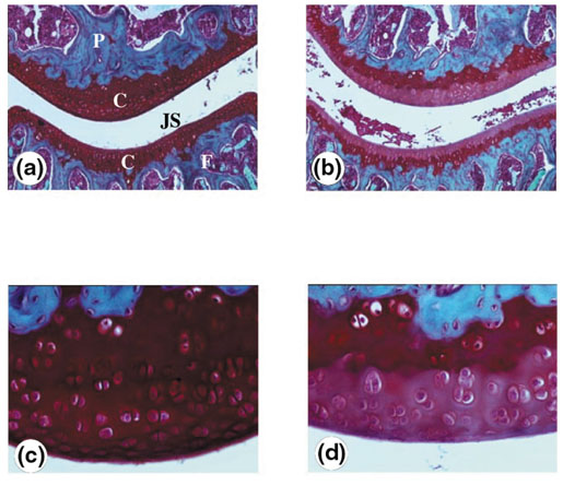
The origin and role of IL-17, a T-cell derived cytokine, in cartilage and bone destruction during rheumatoid arthritis (RA) remain to be clarified. In human ex vivo models, addition of IL-17 enhanced IL-6 production and collagen destruction, and inhibited collagen synthesis by RA synovium explants. On mouse cartilage, IL-17 enhanced cartilage proteoglycan loss and inhibited its synthesis. On human RA bone explants, IL-17 also increased bone resorption and decreased formation. Addition of IL-1 in these conditions increased the effect of IL-17. Blocking of bone-derived endogenous IL-17 with specific inhibitors resulted in a protective inhibition of bone destruction. Conversely, intra-articular administration of IL-17 into a normal mouse joint induced cartilage degradation. In conclusion, the contribution of IL-17 derived from synovium and bone marrow T cells to joint destruction suggests the control of IL-17 for the treatment of RA.


Figures

Figure 1
Figure 1
Effect of exogenous IL-17 on IL-6 production by RA synovium. Synovium samples from RA patients were incubated for 7 days in the presence of IL-17 (50 ng/ml; n = 10), IL-1 (100 pg/ml; n = 10), and IL-17 + IL-1 (n = 3). ELISA measured IL-6 levels in supernatants. Results are expressed as mean ± SEM of % induction of IL-6 production. Spontaneous production of IL-6 was 202 ± 57 ng/ml. *P < 0.01, **P < 0.05 compared with control (medium alone).
Figure 2
Figure 2
Effect of exogenous IL-17 on type I collagen metabolism in RA synovium explants. Synovium samples from RA patients were incubated for 7 days in the presence of IL-17 (50 ng/ml) or IL-1 (100 pg/ml). (a) PICP (n = 4) levels in supernatants were measured by ELISA. Results are expressed as mean ± SEM of % induction of PICP production. Spontaneous production of PICP was 371 ± 36 ng/ml. (b) CTX levels (n = 7) in supernatants were measured by ELISA. Results are expressed as mean ± SEM. Spontaneous production of CTX was 33 ± 11 ng/ml. *P < 0.05, **P < 0.01 compared with control (medium alone).
Figure 3
Figure 3
Effect of exogenous IL-17 on mouse cartilage proteoglycan breakdown. Cartilage explants of patellae were pulse-labeled with 35S-sulfate for 3 h and incubated with IGF-1 (0.25 μg/ml), with or without IL-17 (10 or 100 ng/ml) and/or IL-1 (10 ng/ml) for 48 h. Cultures were performed with six patellae per variable, and data represent percentages of sulfate incorporation into proteoglycan ± SD relative to the values found with IGF-1 alone.
Figure 4
Figure 4
Effect of exogenous IL-17 on IL-6 production by RA bone explants. Bone samples from RA patients were incubated for 7 days in the presence of IL-17 (50 ng/ml; n = 5), IL-1 (100 pg/ml; n = 5), and IL-17 + IL-1 (n = 2). ELISA measured IL-6 levels in supernatants. Results are expressed as mean ± SEM of % induction of IL-6 production. Spontaneous production of IL-6 was 220 ± 95 ng/ml. *P < 0.05 compared with control (medium alone).
Figure 5
Figure 5
Effect of exogenous IL-17 on type I collagen metabolism by RA bone explants. Bone samples from RA patients were incubated for 7 days in the presence of IL-17 (50 ng/ml) and/or IL-1 (100 pg/ml). (a) PICP levels (n = 3) in supernatants were measured by ELISA. Results are expressed as mean ± SEM of % induction of PICP production. Spontaneous production of PICP was 433 ± 133 ng/ml. (b) CTX levels in supernatants were measured by ELISA. Bone samples were incubated with IL-17 (n = 5), IL-1 (n = 5), and IL-17 + IL-1 (n = 2). Results are expressed as mean ± SEM. Spontaneous production of CTX was 104 ± 55 ng/ml. *P < 0.05 compared with control (medium alone).
Figure 6
Figure 6
Effect of blockade of endogenous IL-17 on synovium and bone destruction. (a) Synovium (n = 5) and (b) bone explants (n = 3) from RA patients were incubated for 7 days with and without blocking anti-IL-17 mAb (10 μg/ml) or sIL-17R (1 μg/ml), which was added at the onset of the culture. CTX levels after 7 days of culture were measured by ELISA. Results are expressed as mean ± SEM of culture triplicates. Differences between IL-17 inhibitor treated groups and control groups were analyzed with the nonparametric Wilcoxon paired t test.*P < 0.05.
Figure 7
Figure 7
Respective contribution of endogenous IL-17 to synovium and bone destruction. Paired bone and synovium pieces from the same joint in two RA patients were cultured for 7 days in the presence of sIL-17R (1 μg/ml). CTX production in supernatants was measured by ELISA, and results are expressed in nanomoles.
Figure 8
Figure 8
Effect of intra-articular administration of IL-17 on cartilage destruction. Naive C57Bl/6 mice were intra-articulately injected into the knee joint with recombinant murine IL-17 (100 ng/ml) at days 0 and 3 (b and d). The contralateral joint received an equal volume (6 μl) of saline (a and c). Knee joints were taken for histology 2 days after the last injection. Proteoglycan depletion was analyzed using Safranin O staining. Cartilage depletion was visualized by diminished staining of the matrix. The effect of IL-17 can be seen as the lighter (b and d) rather than the darker staining (a and c) in cartilage. (a) and (b), Original magnification, × 50; (c) and (d), original magnification × 200. P, Patella; F, femur; JS, joint space; C, cartilage.

References

    1. Arend WP. The pathophysiology and treatment of rheumatoid arthritis. Arthritis Rheum. 1997;40:595–597. - PubMed
    1. Chizzolini C, Chicheportiche R, Burger D, Dayer JM. Human Th1 cells preferentially induce interleukin (IL)-1beta while Th2 cells induce IL-1 receptor antagonist production upon cell/cell contact with monocytes. Eur J Immunol. 1997;27:171–177. - PubMed
    1. Chomarat P, Rissoan MC, Pin JJ, Banchereau J, Miossec P. Contribution of IL-1, CD 14, CD 13 in the increased IL-6 production during monocyte synoviocyte interactions. J Immunol. 1995;155:3645–3652. - PubMed
    1. Manolagas SC. Role of cytokines in bone resorption. Bone. 1995;17:63S–67S. - PubMed
    1. Lader CS, Flanagan AM. Prostaglandin E2, interleukin-1a, and tumor necrosis factor-α, increase human osteoblast formation and bone resorption in vitro. Endocrinology. 1998;139:3157–3164. - PubMed

Publication types

MeSH terms