

Guard cell volume and pressure measured concurrently by confocal microscopy and the cell pressure probe
- PMID: 11299339
- PMCID: PMC88815
- DOI: 10.1104/pp.125.4.1577
Guard cell volume and pressure measured concurrently by confocal microscopy and the cell pressure probe
Abstract
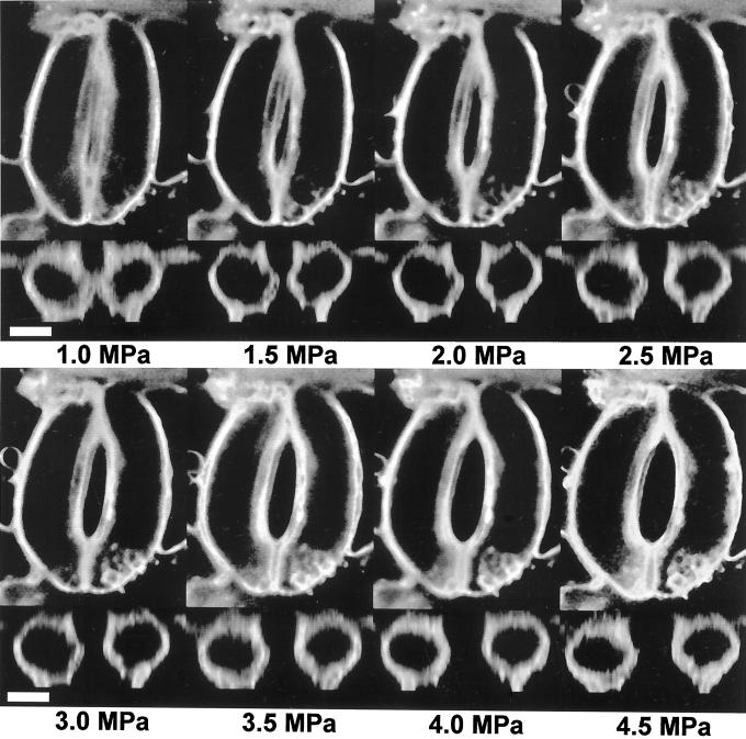
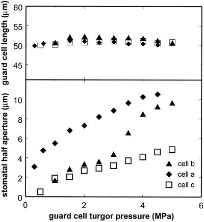
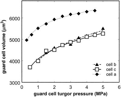
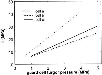
Guard cell turgor pressures in epidermal peels of broad bean (Vicia faba) were measured and controlled with a pressure probe. At the same time, images of the guard cell were acquired using confocal microscopy. To obtain a clear image of guard cell volume, a fluorescent dye that labels the plasma membrane was added to the solution bathing the epidermal peel. At each pressure, 17 to 20 optical sections (each 2 microm thick) were acquired. Out-of-focus light in these images was removed using blind deconvolution, and volume was estimated using direct linear integration. As pressure was increased from as low as 0.3 MPa to as high as 5.0 MPa, guard cell volume increased in a saturating fashion. The elastic modulus was calculated from these data and was found to range from approximately 2 to 40 MPa. The data allow inference of guard cell osmotic content from stomatal aperture and facilitate accurate mechanistic modeling of epidermal water relations and stomatal functioning.
Figures
References
-
- Edwards M, Meidner H, Sheriff DW. Direct measurement of turgor pressure potentials of guard cells: II. The mechanical advantage of subsidiary cells, the spannungsphase, and the optimum leaf water deficit. J Exp Bot. 1976;27:163–171.
-
- Franks PJ, Cowan IR, Farquhar GD. A study of stomatal mechanics using the cell pressure probe. Plant Cell Environ. 1998;21:94–100.
-
- Franks PJ, Cowan IR, Tyerman SD, Cleary AL, Lloyd J, Farquhar GD. Guard cell pressure/aperture characteristics measured with the pressure probe. Plant Cell Environ. 1995;18:795–800.
-
- Fricker M, White N. Volume measurements of guard cell vacuoles during stomatal movements using confocal microscopy. Trans R Microsc Soc. 1990;1:345–348.
Publication types
MeSH terms
LinkOut - more resources
Full Text Sources
Research Materials
