. 2001 Apr;125(4):1611-9.
doi: 10.1104/pp.125.4.1611.

Association of spectrin-like proteins with the actin-organized aggregate of endoplasmic reticulum in the Spitzenkörper of gravitropically tip-growing plant cells

Affiliations

Association of spectrin-like proteins with the actin-organized aggregate of endoplasmic reticulum in the Spitzenkörper of gravitropically tip-growing plant cells

M Braun. Plant Physiol. 2001 Apr.

Abstract

Spectrin-like epitopes were immunochemically detected and immunofluorescently localized in gravitropically tip-growing rhizoids and protonemata of characean algae. Antiserum against spectrin from chicken erythrocytes showed cross-reactivity with rhizoid proteins at molecular masses of about 170 and 195 kD. Confocal microscopy revealed a distinct spherical labeling of spectrin-like proteins in the apices of both cell types tightly associated with an apical actin array and a specific subdomain of endoplasmic reticulum (ER), the ER aggregate. The presence of spectrin-like epitopes, the ER aggregate, and the actin cytoskeleton are strictly correlated with active tip growth. Application of cytochalasin D and A23187 has shown that interfering with actin or with the calcium gradient, which cause the disintegration of the ER aggregate and abolish tip growth, inhibits labeling of spectrin-like proteins. At the beginning of the graviresponse in rhizoids the labeling of spectrin-like proteins remained in its symmetrical position at the cell tip, but was clearly displaced to the upper flank in gravistimulated protonemata. These findings support the hypothesis that a displacement of the Spitzenkörper is required for the negative gravitropic response in protonemata, but not for the positive gravitropic response in rhizoids. It is evident that the actin/spectrin system plays a role in maintaining the organization of the ER aggregate and represents an essential part in the mechanism of gravitropic tip growth.


Figures
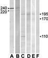
Figure 1
Figure 1
Western blots (7.5% [w/v] SDS gel) of human erythrocyte spectrin (A and B) and a protein extract of Chara rhizoids (C–F). For immunodetection, antibodies raised against human erythrocyte spectrin (A and C) and chicken erythrocyte spectrin (B and D–F) were used. In A and B, the antibodies recognized α- and β-spectrin at 220 and 240 kD, whereas in the rhizoid extract, the anti-spectrin antibodies detected bands at 170 and 195 kD. The use of homogenization buffer that contained 0.1% (w/v) Triton X-100 (D) resulted in a slightly stronger staining of the bands (compare with E). Pre-absorbing anti-chicken antibodies with human spectrin strongly reduced staining of the bands (F).
Figure 2
Figure 2
Localization of actin and spectrin-like epitopes in the apex of a Chara rhizoid (A–D) and a Chara protonema (E–H) by immunofluorescence double labeling. A and E, Labeling with anti-actin; B and F, labeling with anti-chicken spectrin; C and G, overlay of A and B, E and F, respectively. The position of the ER aggregate indicated by the spherical clear zone in the corresponding differential interference contrast (DIC) image (D and H) is marked by the arrows in each image. Optical section images of 1-μm thickness. Bar = 5 μm.
Figure 3
Figure 3
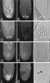
A through C, Immunofluorescence double labeling of a rhizoid with anti-actin (A) and with immunodepleted anti-spectrin (B). The position of the ER aggregate (arrows) is recognizable as a spherical clear zone in the DIC image (C) and in the form of a dense actin array (A), but is only faintly visualized by immunodepleted anti-spectrin (B). Projections of five serial images taken at 0.8-μm z-steps. D through F, Immunofluorescence labeling of a rhizoid with anti-actin (D) and with rabbit serum replacing anti-spectrin (E). The position of the ER aggregate (arrows) is recognizable in the DIC image (F) and in D, but no specific labeling was produced by the rabbit serum (E). Projections of three serial images taken at 0.8-μm z-steps. G through I, Imunofluorescence labeling of a rhizoid with anti-actin (G) and anti-spectrin (H) after application of 10 μm cytochalasin D for 15 min. The ER aggregate has become disintegrated and is no longer recognizable in the DIC image (I), actin microfilament bundles are strongly fragmented (G), and no spectrin-like epitopes are labeled (H). Projections of five serial images taken at 1-μm z-steps. J through L, Immunofluorescence labeling of a rhizoid, which was treated with 2 μm A23187 for 10 min, with anti-actin (J) and anti-spectrin (K). The ER aggregate is not visible in the DIC image (L). Actin microfilaments form thick, randomly oriented bundles in the apex and appear fragmented in the subapical zone (J). Spectrin-like epitopes are not detected (K). Projections of five serial images taken at 1-μm z-steps. Bars = 5 μm.
Figure 4
Figure 4
Graph showing the rates of elongation growth of a representative Chara rhizoid prior to and after incubation with 2 μm A23187 for 10 min (area of lighter gray color) and the corresponding spectrin-immunolabeling images (A–D). The result of spectrin immunolabeling is demonstrated before (A) and 30 min after the treatment (B). The reappearance of spectrin-like epitopes (C and D) and the reorganization of the actin cytoskeleton (D') is shown during the formation (C) and outgrowth of the new tip (D and D') after resumption of tip-growth activity. Spectrin fluorescence reappears in the form of a small patch close to the apical membrane, and later resumes its original position and size in the center of the Spitzenkörper. Projections of six serial images taken at 1-μm z-steps.
Figure 5
Figure 5
Electron microscopic images of the apex of an untreated (A) and a A23187-treated Chara rhizoid (B). In the actively tip-growing cell (A), the ER aggregate (arrows) is located in the center of the Spitzenkörper with its abundant vesicles. Incubation with 2 μm A23187 for 15 min caused a dispersion of the ER aggregate and resulted in a random distribution of ER membranes in the rhizoid apex. Bars = 5 μm.
Figure 6
Figure 6
Localization of spectrin-like proteins in a Chara rhizoid (A) and a Chara protonema (B) at the beginning of the opposite graviresponses after 15 min in a horizontal position. A, In the rhizoid, the labeling of spectrin-like proteins, indicating the position of the ER aggregate, is still located close to the growth center at the tip. B, In the protonema, the labeling of spectrin-like proteins is clearly displaced toward the upper flank where the future outgrowth starts with the formation of a bulge. Broken lines outline the outermost tip region and indicate the median line of the cells. Optical section images of 1.2-μm thickness. Bars = 5 μm.

References

    1. Bartnik E, Hejnowicz Z, Sievers A. Shuttle-like movements of Golgi vesicles in the tip of growing Chara rhizoids. Protoplasma. 1990;159:1–8.
    1. Bartnik E, Sievers A. In vivo observation of a spherical aggregate of endoplasmic reticulum and of Golgi vesicles in the tip of fast-growing Chara rhizoids. Planta. 1988;176:1–9. - PubMed
    1. Baumann O. Association of spectrin with a subcompartment of the endoplasmic reticulum in honeybee photoreceptor cells. Cell Motil Cytoskel. 1998;41:74–86. - PubMed
    1. Beck KA, Buchanan JA, Malhotra V, Nelson WJ. Golgi spectrin: identification of an erythroid β-spectrin homolog associated with the Golgi complex. J Cell Biol. 1994;127:707–723. - PMC - PubMed
    1. Beck KA, Buchanan JA, Nelson WJ. Golgi membrane skeleton: identification, localization and oligomerization of a 195-kDa ankyrin isoform associated with the Golgi complex. J Cell Sci. 1997;110:1239–1249. - PubMed

Publication types

LinkOut - more resources