. 2001 Apr;125(4):1880-90.
doi: 10.1104/pp.125.4.1880.

N-acetylglucosamine and glucosamine-containing arabinogalactan proteins control somatic embryogenesis

Affiliations

N-acetylglucosamine and glucosamine-containing arabinogalactan proteins control somatic embryogenesis

A J van Hengel et al. Plant Physiol. 2001 Apr.

Abstract

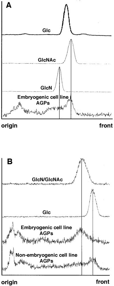
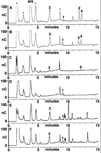
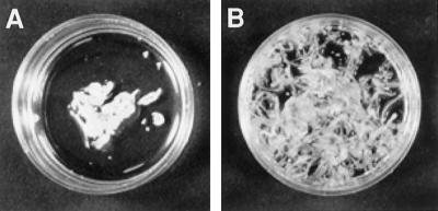
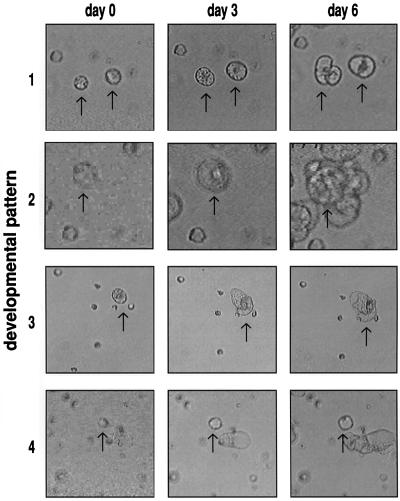
In plants, complete embryos can develop not only from the zygote, but also from somatic cells in tissue culture. How somatic cells undergo the change in fate to become embryogenic is largely unknown. Proteins, secreted into the culture medium such as endochitinases and arabinogalactan proteins (AGPs) are required for somatic embryogenesis. Here we show that carrot (Daucus carota) AGPs can contain glucosamine and N-acetyl-D-glucosaminyl and are sensitive to endochitinase cleavage. To determine the relevance of this observation for embryogenesis, an assay was developed based on the enzymatic removal of the cell wall from cultured cells. The resulting protoplasts had a reduced capacity for somatic embryogenesis, which could be partially restored by adding endochitinases to the protoplasts. AGPs from culture medium or from immature seeds could fully restore or even increase embryogenesis. AGPs pretreated with chitinases were more active than untreated molecules and required an intact carbohydrate constituent for activity. AGPs were only capable of promoting embryogenesis from protoplasts in a short period preceding cell wall reformation. Apart from the increase in embryogenesis, AGPs can reinitiate cell division in a subpopulation of otherwise non-dividing protoplasts. These results show that chitinase-modified AGPs are extracellular matrix molecules able to control or maintain plant cell fate.


Figures

Figure 1
Figure 1
Optical density scans of autoradiograms of TLCs. The scans represent the mobility of the TFA-degraded AGPs of an embryogenic and a nonembryogenic cell line labeled with 10−7 M 14C]GlcNAc, compared with the mobility of d-[1-14C]labeled Glc, GlcN, and GlcNAc references. A, AGPs and references degraded by means of incubation in 2 m TFA at 100°C for 45 min. B, AGPs and references degraded by means of incubation in 2 m TFA at 120°C for 60 min.
Figure 2
Figure 2
HPAE-PAD chromatography of AGP-derived oligosaccharides. HPAE-PAD chromatograms of AGPs that were isolated from immature seeds treated with pectinase and then incubated with AGP degrading hydrolases in the presence or absence of EP3 endochitinase. Oligosaccharides that change in relative amount upon incubation with EP3 endochitinase are numbered as 1 (retention time of 9 min 10 s), 2 (retention time of 11 min 50 s), 3 (retention time of 7 min), and 4 (retention time of 12 min 20 s). Peak height is expressed in nanocoulomb (nC). A, AGPs incubated with endogalactosidase, endo-, and exoarabinofuranosidase. B, AGPs incubated with endogalactosidase, endo-, and exoarabinofuranosidase in combination with EP3 endochitinase. C, AGPs incubated with exoarabinofuranosidase. D, AGPs incubated with exoarabinofuranosidase in combination with EP3 endochitinase. E, AGPs incubated with endoarabinofuranosidase. F, AGPs incubated with endoarabinofuranosidase in combination with EP3 endochitinase.
Figure 3
Figure 3
Restoration of somatic embryogenesis from protoplasts by seed AGPs. A, Callus and an occasional somatic embryo formed in unsupplemented control culture. B, Plantlets formed after addition of 3 μg mL−1 of immature 21 DAP seed AGPs, pretreated with pectinase.
Figure 4
Figure 4
Development of immobilized carrot protoplasts. Development of individual carrot protoplasts was analyzed by means of video cell tracking. After comparison of images as obtained after 0, 3, and 6 d, four patterns of development were identified: 1, cells that only divide and do not enlarge; 2, cells that enlarge and divide; 3, cells that only enlarge; and 4, cells that do not divide or enlarge. Arrows indicate cells representing the developmental pattern indicated.

References

    1. Aitkin M, Anderson D, Francis B. Statistical Modelling in GLIM. Oxford: Oxford University Press; 1991.
    1. Bacic A, Harris PJ, Stone BA. Structure and function of plant cell walls. In: Preiss J, editor. The Biochemistry of Plants: a Comprehensive Treatise. San Diego: Academic Press; 1988. pp. 297–371.
    1. Baldan B, Guzzo F, Filippini F, Gasparian M, LoSchiavo F, Vitale A, De Vries SC, Mariani P, Terzi M. The secretory nature of the lesion of carrot cell variant ts11, rescuable by endochitinase. Planta. 1997;203:381–389. - PubMed
    1. Baldwin TC, McCann MC, Roberts K. A novel hydroxyproline-deficient arabinogalactan protein secreted by suspension-cultured cells of Daucus carota: purification and partial characterization. Plant Physiol. 1993;103:115–123. - PMC - PubMed
    1. Brown JA, Fry SC. Novel O-d-galacturonyl esters in the pectic polysaccharides of suspension-cultured plant cells. Plant Physiol. 1993;10:993–999. - PMC - PubMed

Publication types

LinkOut - more resources