. 2001 Apr;125(4):2040-52.
doi: 10.1104/pp.125.4.2040.

Pollen tubes of Nicotiana alata express two genes from different beta-glucan synthase families

Affiliations

Pollen tubes of Nicotiana alata express two genes from different beta-glucan synthase families

M S Doblin et al. Plant Physiol. 2001 Apr.

Abstract

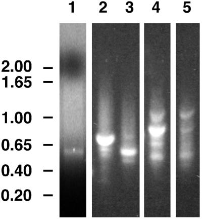
The walls deposited by growing pollen tubes contain two types of beta-glucan, the (1,3)-beta-glucan callose and the (1,4)-beta-glucan cellulose, as well as various alpha-linked pectic polysaccharides. Pollen tubes of Nicotiana alata Link et Otto, an ornamental tobacco, were therefore used to identify genes potentially encoding catalytic subunits of the callose synthase and cellulose synthase enzymes. Reverse transcriptase-polymerase chain reactions (RT-PCR) with pollen-tube RNA and primers designed to conserved regions of bacterial and plant cellulose synthase (CesA) genes amplified a fragment that corresponded to an abundantly expressed cellulose-synthase-like gene named NaCslD1. A fragment from a true CesA gene (NaCesA1) was also amplified, but corresponding cDNAs could not be identified in a pollen-tube library, consistent with the very low level of expression of the NaCesA1 gene. RT-PCR with pollen-tube RNA and primers designed to regions conserved between the fungal FKS genes [that encode (1,3)-beta-glucan synthases] and their presumed plant homologs (the Gsl or glucan-synthase-like genes) amplified a fragment that corresponded to an abundantly expressed gene named NaGsl1. A second Gsl gene detected by RT-PCR (NaGsl2) was expressed at low levels in immature floral organs. The structure of full-length cDNAs of NaCslD1, NaCesA1, and NaGsl1 are presented. Both NaCslD1 and NaGsl1 are predominantly expressed in the male gametophyte (developing and mature pollen and growing pollen tubes), and we propose that they encode the catalytic subunits of two beta-glucan synthases involved in pollen-tube wall synthesis. Different beta-glucans deposited in one cell type may therefore be synthesized by enzymes from different gene families.


Figures

Figure 1
Figure 1
RT-PCR amplification of fragments encoding putative β-glucan synthases. Lanes 1 through 3 show products of reactions containing primers UGF and DOMB1R for (1,4)-β-glucan synthases. Lanes 4 and 5 show products of reactions containing primers Fks3F and Fks7R for (1,3)-β-glucan synthases. Lane 1 (control) used the rice CesA EST D47622 as template; lanes 2 and 4 used cDNA from N. alata pollen tubes grown in vitro for 12 h as template; and lanes 3 and 5 used cDNA from young expanding N. alata leaves as template. Numbers to the left show fragment sizes in kilobase pairs.
Figure 2
Figure 2
Distance cladogram for polypeptide sequences deduced from CesA and CslD genes. Three A. xylinum CesA sequences (AcsAB, BcsA, and AcsAII; Wong et al., 1990; Saxena and Brown, 1995) were included as an outgroup. Bootstrap supports are indicated along the relevant branches.
Figure 3
Figure 3
Domain structure and topology of the predicted NaCslD1 protein. A, Predicted NaCslD1 protein with predicted AtCesA1/RSW1 protein (Arioli et al., 1998) as comparison, drawn to scale as boxes. Length of total amino acid sequence is shown at the end of each box. Proteins are divided into the seven domains defined by Pear et al. (1996). H-1, H-2, and H-3 (gray boxes) are homology domains; P-CR, plant conserved region; HVR, hypervariable region; N and C refer to the N- and C-terminal domains, respectively. Numbers above the NaCslD1 box and below the AtCesA1 box show the number of amino acids within each domain. Amino acid identity (%) within a domain is given below the figure. Black boxes define the regions containing motifs of the conserved D,D,D,QXXRW domain as defined by Pear et al. (1996). A hatched box indicates the Cys-rich LIM domain. Dashed boxes in the NaCslD1 sequence indicate the insertion in the N-terminal domain, and the three insertions in the P-CR region. The position corresponding to the 700-bp UGF-DOMB1R fragment produced by RT-PCR is also shown. B, Hydropathy plot of the predicted NaCslD1 protein. Horizontal black bars indicate the eight predicted TMHs. C, Topology model of NaCslD1 based on the GhCesA1 model proposed by Pear et al. (1996). Cw, Cell wall; PM, plasma-membrane; cyt, cytoplasm; ●, location of potential N-glycosylation sites; 1 through 8, the TMHs.
Figure 4
Figure 4
Distance cladogram for polypeptide sequences deduced from plant Gsl genes. Five fungal FKS sequences (ScFks1, AfFks1, FnFKS1, PbFKS1, and CaGSC1; Douglas et al., 1994; Kelly et al., 1996) were included as an outgroup. Bootstrap supports are indicated along the relevant branches.
Figure 5
Figure 5
Domain structure and topology of the predicted NaGsl1 protein. A, Predicted NaGsl1 protein, with predicted CFL1 (Cui et al., 1999) as comparison, drawn to scale as boxes. Length of total amino acid sequence is shown at the end of each box. Proteins are divided into the four domains defined by Douglas et al. (1994). N, N-terminal domain; TM1 and TM2, first and second transmembrane domains; cyt, cytoplasmic domain. Numbers above the NaGsl1 box and below the CFL1 box show the number of amino acids within each domain. Amino acid identity (%) within a domain is given below the figure. The position corresponding to the 820-bp Fks3F-7R fragment produced by RT-PCR is also shown. B, Hydropathy plot of the predicted NaGsl1 protein. Horizontal black bars indicate the 16 TMHs. C, Topology model of NaCslD1 based on the yeast FKS1 model (Douglas et al., 1994). 1 through 16, The tMHs. Abbreviations and locations of potential N-glycosylation sites are as for Figure 3.
Figure 6
Figure 6
RNA-blot analysis of NaCslD1, NaCesA1, NaGsl1, and NaGsl2 expression. N. alata tissues: SE, sepal; PE, petal; AN, anther; ST, style; OV, ovary; MP, mature pollen; 4 through 24, pollen tubes grown for various times, given as h; L, leaf; R, root; S, stem; LS, light-grown seedling; and DS, dark-grown seedling. N. plumbaginifolia tissues: CC, suspension-cultured cells. Transcript sizes are indicated on the right of the figure. A, Blots probed with DIG-labeled UGF-DOMB1R fragment of NaCslD1. Flower buds and vegetative tissues: hybridized and washed at 35°C, exposed for 2 min. Pollen and pollen tubes: hybridized and washed at 42°C, exposed for 2 min. B, Flower buds, pollen, and pollen tubes: blots probed with 32P-labeled UGF-DOMB1R fragment of NaCesA1, hybridized, and washed at 42°C, exposed for 21 d. Vegetative tissues: blots probed with DIG-labeled UGF-DOMB1R fragment of NaCesA1, hybridized, and washed at 37°C, exposed for 2 h. C, Blots probed with DIG-labeled Fks3F-Fks7R fragment of NaGsl1. Flower buds, pollen, pollen tubes, and vegetative tissues: hybridized at 42°C and washed at 65°C, exposed for 1.5 h. D, Blots probed with DIG-labeled Fks3F-Fks7R fragment of NaGsl2. Flower buds, pollen, pollen tubes, and vegetative tissues: hybridized at 42°C, washed at 65°C, and exposed for 1.5 h.

References

    1. Antelo L, Cosio EG, Hertkorn N, Ebel J. Partial purification of a GTP-insensitive (1,3)-β-glucan synthase from Phytophthora sojae. FEBS Lett. 1998;433:191–195. - PubMed
    1. Arioli T, Peng L, Betzner AS, Burn J, Wittke W, Herth W, Camilleri C, Höfte H, Plazinski J, Birch R. Molecular analysis of cellulose biosynthesis in Arabidopsis. Science. 1998;279:717–720. - PubMed
    1. Bacic A, Harris PJ, Stone BA. Structure and function of plant cell walls. In: Priess J, editor. The Biochemistry of Plants. New York: Academic Press; 1988. pp. 297–371.
    1. Becker M, Vincent C, Reid JSG. Biosynthesis of (1,3)(1,4)-β-glucan and (1,3)-β-glucan in barley (Hordeum vulgare L): properties of the membrane-bound glucan synthases. Planta. 1995;195:331–338. - PubMed
    1. Billongrand C, Marais MF, Joseleau JP, Girard V, Gay L, Fevre M. A novel 1,3-β-glucan synthase from the oomycete Saprolegnia monoica. Microbiology. 1997;143:3175–3187. - PubMed

Publication types

MeSH terms

LinkOut - more resources