

Role of mast cells in chronic stress induced colonic epithelial barrier dysfunction in the rat
- PMID: 11302959
- PMCID: PMC1728291
- DOI: 10.1136/gut.48.5.630
Role of mast cells in chronic stress induced colonic epithelial barrier dysfunction in the rat
Abstract
Background and aims: Stress may be an important factor in exacerbating inflammatory bowel disease but the underlying mechanism is unclear. Defective epithelial barrier function may allow uptake of luminal antigens that stimulate an immune/inflammatory response. Here, we examined the effect of chronic stress on colonic permeability and the participation of mast cells in this response.
Methods: Mast cell deficient Ws/Ws rats and +/+ littermate controls were submitted to water avoidance stress or sham stress (one hour/day) for five days. Colonic epithelial permeability to a model macromolecular antigen, horseradish peroxidase, was measured in Ussing chambers. Epithelial and mast cell morphology was studied by light and electron microscopy.
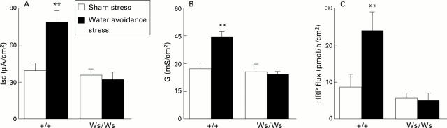
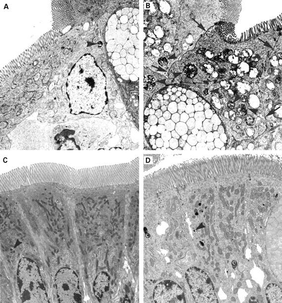
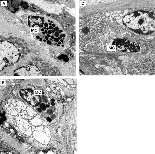
Results: Chronic stress significantly increased macromolecular flux and caused epithelial mitochondrial swelling in +/+ rats, but not in Ws/Ws rats, compared with non-stressed controls. Stress increased the number of mucosal mast cells and the proportion of cells showing signs of activation in +/+ rats. No mast cells or ultrastructural abnormalities of the epithelium were present in Ws/Ws rats. Increased permeability in +/+ rats persisted for 72 hours after stress cessation.
Conclusions: Chronic stress causes an epithelial barrier defect and epithelial mitochondrial damage, in parallel with mucosal mast cell hyperplasia and activation. The study provides further support for an important role for mast cells in stress induced colonic mucosal pathophysiology.
Figures
Similar articles
-
Chronic stress impairs rat growth and jejunal epithelial barrier function: role of mast cells.Am J Physiol Gastrointest Liver Physiol. 2000 Jun;278(6):G847-54. doi: 10.1152/ajpgi.2000.278.6.G847. Am J Physiol Gastrointest Liver Physiol. 2000. PMID: 10859213
-
Chronic peripheral administration of corticotropin-releasing factor causes colonic barrier dysfunction similar to psychological stress.Am J Physiol Gastrointest Liver Physiol. 2008 Sep;295(3):G452-9. doi: 10.1152/ajpgi.90210.2008. Epub 2008 Jul 17. Am J Physiol Gastrointest Liver Physiol. 2008. PMID: 18635602
-
Chronic stress induces mast cell-dependent bacterial adherence and initiates mucosal inflammation in rat intestine.Gastroenterology. 2002 Oct;123(4):1099-108. doi: 10.1053/gast.2002.36019. Gastroenterology. 2002. PMID: 12360472
-
Intestinal Mucosal Mast Cells: Key Modulators of Barrier Function and Homeostasis.Cells. 2019 Feb 8;8(2):135. doi: 10.3390/cells8020135. Cells. 2019. PMID: 30744042 Free PMC article. Review.
-
Review article: mechanisms of initiation and perpetuation of gut inflammation by stress.Aliment Pharmacol Ther. 2002 Dec;16(12):2017-28. doi: 10.1046/j.1365-2036.2002.01359.x. Aliment Pharmacol Ther. 2002. PMID: 12452934 Review.
Cited by
-
Stereotaxic Exposure of the Central Nucleus of the Amygdala to Corticosterone Increases Colonic Permeability and Reduces Nerve-Mediated Active Ion Transport in Rats.Front Neurosci. 2018 Aug 7;12:543. doi: 10.3389/fnins.2018.00543. eCollection 2018. Front Neurosci. 2018. PMID: 30154689 Free PMC article.
-
The role of mast cells in pediatric gastrointestinal disease.Ann Gastroenterol. 2019 Jul-Aug;32(4):338-345. doi: 10.20524/aog.2019.0378. Epub 2019 Apr 22. Ann Gastroenterol. 2019. PMID: 31263355 Free PMC article. Review.
-
Loss of Bladder Epithelium Induced by Cytolytic Mast Cell Granules.Immunity. 2016 Dec 20;45(6):1258-1269. doi: 10.1016/j.immuni.2016.11.003. Epub 2016 Dec 6. Immunity. 2016. PMID: 27939674 Free PMC article.
-
Mind-altering microorganisms: the impact of the gut microbiota on brain and behaviour.Nat Rev Neurosci. 2012 Oct;13(10):701-12. doi: 10.1038/nrn3346. Epub 2012 Sep 12. Nat Rev Neurosci. 2012. PMID: 22968153 Review.
-
The Gut-Brain Axis, BDNF, NMDA and CNS Disorders.Neurochem Res. 2016 Nov;41(11):2819-2835. doi: 10.1007/s11064-016-2039-1. Epub 2016 Aug 23. Neurochem Res. 2016. PMID: 27553784 Review.
References
Publication types
MeSH terms
Substances
LinkOut - more resources
Full Text Sources
Other Literature Sources
Medical