

Effect of power Doppler and digital subtraction techniques on the comparison of myocardial contrast echocardiography with SPECT
- PMID: 11303008
- PMCID: PMC1729711
- DOI: 10.1136/heart.85.5.549
Effect of power Doppler and digital subtraction techniques on the comparison of myocardial contrast echocardiography with SPECT
Abstract
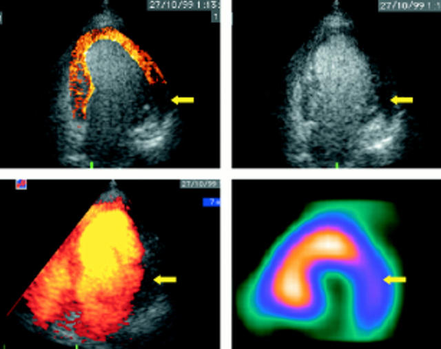
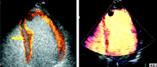
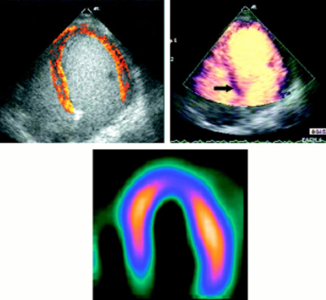
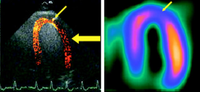
Objective: To compare the accuracy and feasibility of harmonic power Doppler and digitally subtracted colour coded grey scale imaging for the assessment of perfusion defect severity by single photon emission computed tomography (SPECT) in an unselected group of patients.
Design: Cohort study.
Setting: Regional cardiothoracic unit.
Patients: 49 patients (mean (SD) age 61 (11) years; 27 women, 22 men) with known or suspected coronary artery disease were studied with simultaneous myocardial contrast echo (MCE) and SPECT after standard dipyridamole stress.
Main outcome measures: Regional myocardial perfusion by SPECT, performed with (99m)Tc tetrafosmin, scored qualitatively and also quantitated as per cent maximum activity.
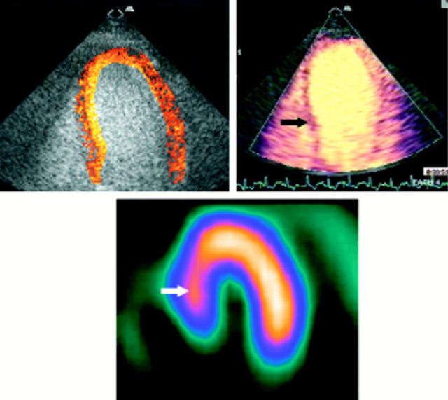
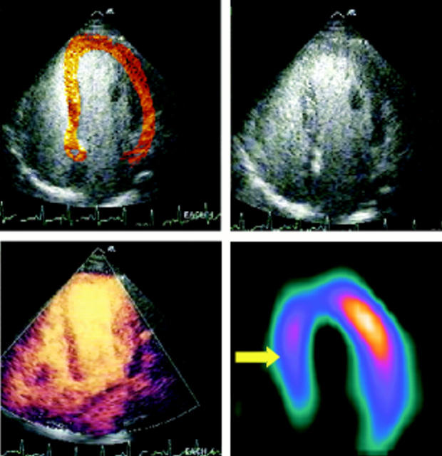
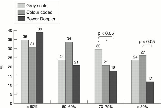
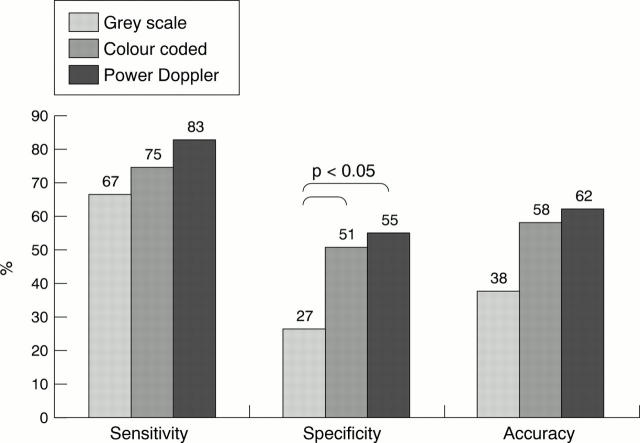
Results: Normal perfusion was identified by SPECT in 225 of 270 segments (83%). Contrast echo images were interpretable in 92% of patients. The proportion of normal MCE by grey scale, subtracted, and power Doppler techniques were respectively 76%, 74%, and 88% (p < 0.05) at > 80% of maximum counts, compared with 65%, 69%, and 61% at < 60% of maximum counts. For each technique, specificity was lowest in the lateral wall, although power Doppler was the least affected. Grey scale and subtraction techniques were least accurate in the septal wall, but power Doppler showed particular problems in the apex. On a per patient analysis, the sensitivity was 67%, 75%, and 83% for detection of coronary artery disease using grey scale, colour coded, and power Doppler, respectively, with a significant difference between power Doppler and grey scale only (p < 0.05). Specificity was also the highest for power Doppler, at 55%, but not significantly different from subtracted colour coded images.
Conclusions: Myocardial contrast echo using harmonic power Doppler has greater accuracy than with grey scale imaging and digital subtraction. However, power Doppler appears to be less sensitive for mild perfusion defects.
Figures
Similar articles
-
Non-invasive detection of coronary artery stenosis: a comparison among power-Doppler contrast echo, 99Tc-Sestamibi SPECT and echo wall-motion analysis.Coron Artery Dis. 2003 May;14(3):239-45. doi: 10.1097/01.mca.0000065924.30342.38. Coron Artery Dis. 2003. PMID: 12702928
-
Detection of myocardial perfusion abnormalities after a recent acute coronary syndrome by quantitative Levovist myocardial contrast echocardiography: comparison with 99m Tc-Myoview SPECT imaging.Can J Cardiol. 2003 Mar 15;19(3):251-6. Can J Cardiol. 2003. PMID: 12677280
-
Assessment of myocardial perfusion with intravenous contrast echocardiography: comparison with (99) Tc-tetrofosmin single photon emission computed tomography and dobutamine echocardiography.Echocardiography. 2003 Jan;20(1):37-45. doi: 10.1046/j.1540-8175.2003.00005.x. Echocardiography. 2003. PMID: 12848696
-
Clinical experience with SonoVue in myocardial perfusion imaging.Echocardiography. 2000 Aug;17(6 Pt 2):S17-23. doi: 10.1111/j.1540-8175.2000.tb01190.x. Echocardiography. 2000. PMID: 11058235 Review.
-
Instrumentation for contrast echocardiography.Echocardiography. 2002 Apr;19(3):241-58. doi: 10.1046/j.1540-8175.2002.00241.x. Echocardiography. 2002. PMID: 12022934 Review.
Cited by
-
Assessment of infarcted myocardium with real time myocardial contrast echocardiography: comparison with technetium-99m sestamibi single photon emission computed tomography.Heart. 2005 Dec;91(12):1568-72. doi: 10.1136/hrt.2004.057844. Epub 2005 Mar 17. Heart. 2005. PMID: 15774606 Free PMC article.
-
Effects of glucose-insulin-potassium infusion on chronic ischaemic left ventricular dysfunction.Heart. 2003 Jan;89(1):61-5. doi: 10.1136/heart.89.1.61. Heart. 2003. PMID: 12482794 Free PMC article.
References
Publication types
MeSH terms
Substances
LinkOut - more resources
Full Text Sources
Miscellaneous