. 2001 Apr 16;193(8):967-74.
doi: 10.1084/jem.193.8.967.

The clinical course of experimental autoimmune encephalomyelitis and inflammation is controlled by the expression of CD40 within the central nervous system

Affiliations

The clinical course of experimental autoimmune encephalomyelitis and inflammation is controlled by the expression of CD40 within the central nervous system

B Becher et al. J Exp Med. .

Abstract

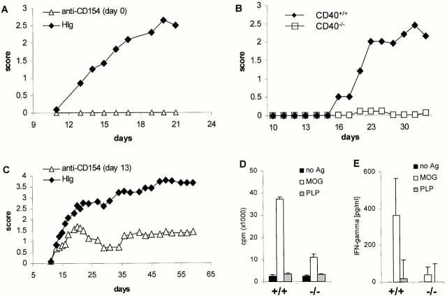
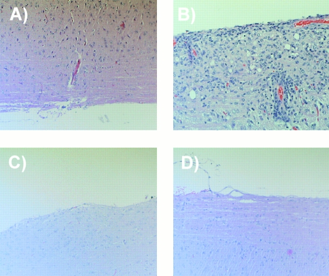
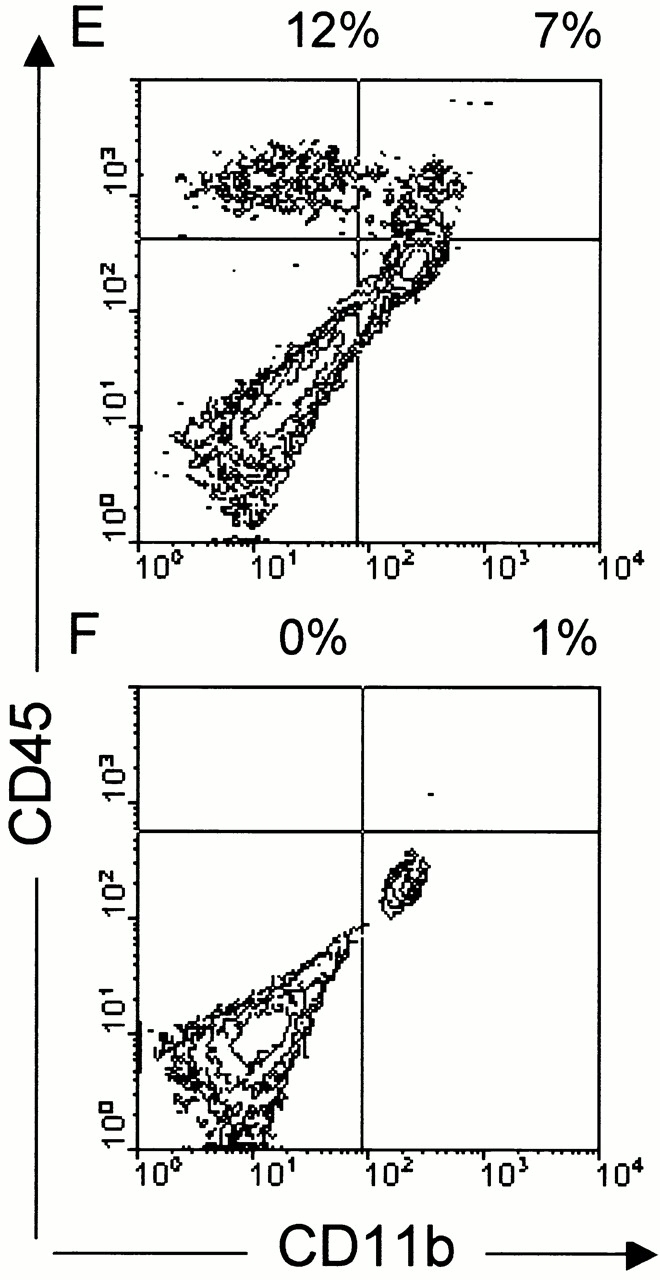
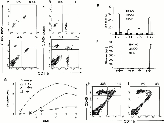
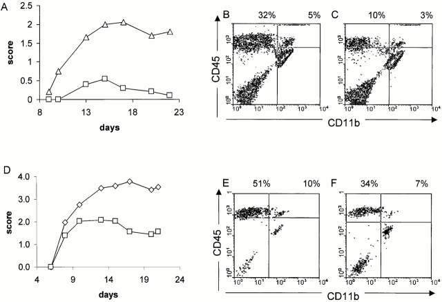
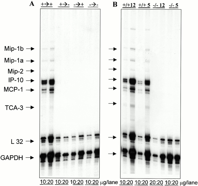
Although it is clear that the function of CD40 on peripheral hematopoietic cells is pivotal to the development of autoimmunity, the function of CD40 in autoimmune disease outside this compartment is unresolved. In a model of experimental autoimmune encephalomyelitis (EAE), evidence is presented that CD40-CD154 interactions within the central nervous system (CNS) are critical determinants of disease development and progression. Using bone marrow (BM) chimeric mice, the data suggest that the lack of expression of CD40 by CNS-resident cells diminishes the intensity and duration of myelin oligodendrocyte glycoprotein (MOG)-induced EAE and also reduces the degree of inflammatory cell infiltrates into the CNS. Although CNS inflammation is compromised in the CD40(+/+)-->CD40(-/-) BM chimeric mice, the restricted CD40 expression had no impact on peripheral T cell priming or recall responses. Analysis of RNA expression levels within the CNS demonstrated that encephalitogenic T cells, which entered a CNS environment in which CD40 was absent from parenchymal microglia, could not elicit the expression of chemokines within the CNS. These data provide evidence that CD40 functions outside of the systemic immune compartment to amplify organ-specific autoimmunity.


Figures

Figure 1
Figure 1
CD40 is critical for the induction of active MOG-EAE in C57BL/6 mice and T cell priming. (A) Early administration of anti-CD154 blocks the development of MOG-induced EAE. Mice were immunized with MOG35–55 in CFA and given 350 μg/mouse of anti-CD154 mAbs three times per week starting at the time of immunization (▵). Control mice were treated with H-Ig mAbs (♦; n = 6/group). (B) CD40−/− mice are not susceptible to MOG-induced EAE. CD40−/− (□) and CD40+/+ (♦) mice were immunized as described in panel A, and disease was monitored (n = 9/group). (C) Treatment of mice with anti-CD154 at disease onset ameliorated MOG-induced EAE. Mice were treated at the first sign of clinical disease with either anti-CD154 (▵) or control H-Ig (♦) (n = 9/group). (D) T cell priming to MOG is reduced in CD40−/− mice. To assess the ability of CD40−/− mice to prime T cells against MOG35–55, CD40+/+ (+/+) or CD40−/− (−/−) mice were immunized with MOG35–55 in CFA. LN cells were harvested after 5 d and stimulated with either no antigen, 100 μg of MOG35–55, or irrelevant PLP peptide. (E) MOG-induced IFN-γ secretion is reduced in CD40−/− mice. IFN-γ production from LN cells primed to MOG35–55 in CD40+/+ and CD40−/− was assessed in vitro as described in D. Displayed is a representative of at least three experiments.
Figure 2
Figure 2
CD40–CD154 blockade inhibits extravasation of leukocytes into the CNS. (A–D) Genetic deficiency of CD40 or anti-CD154 treatment reduces leukocyte infiltration into the CNS as assessed by histochemistry. EAE was induced in either CD40+/+ or CD40−/− mice using MOG35–55 emulsified in CFA as described. CD40+/+ mice were either treated with anti-CD154 or irrelevant H-Ig at the time of immunization (three times per week). To quantify inflammatory infiltration into the CNS, 5–10 mice per group were killed 7 d after disease onset. Spinal cords and cerebella were collected and either fixed in 10% formaldehyde/PBS or homogenized. Spinal cords from: (A) CD40+/+ mice, nonimmune; (B) CD40+/+ mice, immune and treated with H-Ig; (C) CD40−/− mice, immune; and (D) CD40+/+ mice, immune and treated with anti-CD154 at the time of immunization, were sectioned and stained with hematoxylin and eosin. Displayed is a representative of at least three individual experiments. (E and F) Infiltration of leukocyte subsets into the CNS is reduced in CD40−/− mice. Cells derived from spinal cord cell suspension were separated over a discontinuous Percoll gradient and stained with anti-CD45 and anti-CD11b mAbs. Cells were analyzed by flow cytometry; (E) CD40+/+ mice; (F) CD40−/− mice.
Figure 2
Figure 2
CD40–CD154 blockade inhibits extravasation of leukocytes into the CNS. (A–D) Genetic deficiency of CD40 or anti-CD154 treatment reduces leukocyte infiltration into the CNS as assessed by histochemistry. EAE was induced in either CD40+/+ or CD40−/− mice using MOG35–55 emulsified in CFA as described. CD40+/+ mice were either treated with anti-CD154 or irrelevant H-Ig at the time of immunization (three times per week). To quantify inflammatory infiltration into the CNS, 5–10 mice per group were killed 7 d after disease onset. Spinal cords and cerebella were collected and either fixed in 10% formaldehyde/PBS or homogenized. Spinal cords from: (A) CD40+/+ mice, nonimmune; (B) CD40+/+ mice, immune and treated with H-Ig; (C) CD40−/− mice, immune; and (D) CD40+/+ mice, immune and treated with anti-CD154 at the time of immunization, were sectioned and stained with hematoxylin and eosin. Displayed is a representative of at least three individual experiments. (E and F) Infiltration of leukocyte subsets into the CNS is reduced in CD40−/− mice. Cells derived from spinal cord cell suspension were separated over a discontinuous Percoll gradient and stained with anti-CD45 and anti-CD11b mAbs. Cells were analyzed by flow cytometry; (E) CD40+/+ mice; (F) CD40−/− mice.
Figure 3
Figure 3
Analysis of the induction of actively induced EAE in irradiated BM chimeric mice. (A–D) In BM chimeric mice, the parenchymal microglial cells are host derived and the infiltrating leukocytes are predominantly donor derived. Mice were irradiated and reconstituted with CD45.1-congenic BM cells. Engraftment took place over 2 mo. CNS cells of nonimmune mice were stained with either the (A) host (CD45.2) or (B) donor-congenic CD45.1 marker. C and D show the degree of chimerism of CNS-infiltrating leukocytes in BM chimeric mice with EAE induced by immunization with MOG35–55 in CFA. (E and F) The lack of CD40 expression on radioresistant cells in the host does not influence peripheral T cell responses. After 2 mo of recovery, mice were immunized with MOG/CFA and LNs were removed 5 d later. Recall proliferation (E) and IFN-γ secretion (F) in response to either 100 μg MOG peptide or irrelevant PLP peptide was assessed in vitro. (G) The lack of CD40 in the CNS reduces EAE. CD40+/+→CD40+/+, CD40+/+→CD40−/−, CD40−/−→CD40+/+, and CD40−/−→CD40−/− mice were produced, allowed to recover for 2 mo, then immunized with MOG35–55 in CFA as described above and scored for clinical disease (n = 5/group; representative of four individual experiments). The clinical score of CD40+/+→CD40+/+ vs. CD40+/+→CD40−/− is statistically significantly different (P < 0.05). EAE was scored over 24 d. (H and I) The lack of CD40 expression in the CNS reduces the extent of leukocyte infiltration. 7 d after disease onset, leukocyte infiltration into the CNS of (H) CD40+/+→CD40+/+ and (I) CD40+/+→CD40−/− mice was analyzed as described.
Figure 4
Figure 4
Encephalitogenic T cells are less effective at inducing EAE when transferred into a CD40-deficient host. (A) Adoptive transfer of MOG-reactive T cells into a CD40−/− host compared with transfer into CD40+/+ host results in less disease. MOG-reactive cells (15 × 106) were adoptively transferred into either CD40+/+ (▵) or CD40−/− (□) recipients and the mice were scored for clinical disease (n = 6/group). (B and C) Adoptive transfer of MOG-reactive T cells can less effectively infiltrate the CNS in a CD40−/− host. 7 d after disease onset, mice were killed and CNS infiltrates into (B) CD40+/+ and (C) CD40−/− mice were analyzed as described. (D) Adoptive transfer of MOG-reactive T cells into CD40+/+→CD40−/− BM chimeras results in less disease compared with CD40+/+→CD40+/+ mice. MOG-reactive cells (20 × 106) were adoptively transferred into either CD40+/+→CD40+/+ (⋄) or CD40+/+→CD40−/− (□) recipients and the mice were scored for clinical disease (n = 6/group). (E and F) Adoptive transfer of MOG-reactive T cells can less effectively infiltrate when CD40 is absent from the CNS. 7 d after disease onset, mice were killed and CNS infiltrates into (E) CD40+/+→CD40+/+ and (F) CD40+/+→CD40−/− were analyzed as described.
Figure 5
Figure 5
Chemokine expression within the CNS of CD40+/+, CD40−/−, and BM chimeric mice. (A) Chemokine expression is reduced in BM chimeras where CD40 is absent in the CNS. EAE was induced in BM chimeric mice by active immunization (+→+ = CD40+/+ → CD40+/+; +→− = CD40+/+ → CD40−/−; −→+ = CD40−/− →CD40+/+; −→− = CD40−/−→CD40−/−). 5 d after disease onset the animals were killed and total RNA was isolated from spinal cords and analyzed for chemokine expression by RNase protection assay. Either 10 or 20 mg of RNA was loaded onto the gel. (B) Chemokine expression is reduced in CD40−/− mice upon passive transfer of MOG-reactive cells. EAE was induced in either CD40+/+ or CD40−/− mice by adoptive transfer of either 12 × 106 or 5 × 10⁶ MOG-reactive cells and RNase protection assay was performed as described. Mip, macrophage inflammatory protein; IP-10, IFN-γ–inducible protein 10; MCP-1, monocyte chemoattractant protein 1; TCA-3, T cell activation gene 3; GAPDH, glyceraldehyde 3-phosphate dehydrogenase.

Similar articles

Cited by

References

    1. Grewal I.S., Flavell R.A. CD40 and CD154 in cell-mediated immunity. Annu. Rev. Immunol. 1998;16:111–135. - PubMed
    1. Mackey M.F., Barth R.J.J., Noelle R.J. The role of CD40/CD154 interactions in the priming, differentiation, and effector function of helper and cytotoxic T cells. J. Leukoc. Biol. 1998;63:418–428. - PubMed
    1. Durie F.H., Fava R.A., Foy T.M., Aruffo A., Ledbetter J.A., Noelle R.J. Prevention of collagen-induced arthritis with an antibody to gp39, the ligand for CD40. Science. 1993;261:1328–1330. - PubMed
    1. Sadlack B., Lohler J., Schorle H., Klebb G., Haber H., Sickel E., Noelle R.J., Horak I. Generalized autoimmune disease in interleukin-2-deficient mice is triggered by an uncontrolled activation and proliferation of CD4+ T cells. Eur. J. Immunol. 1995;25:3053–3059. - PubMed
    1. Gerritse K., Laman J.D., Noelle R.J., Aruffo A., Ledbetter J.A., Boersma W.J., Claassen E. CD40-CD40 ligand interactions in experimental allergic encephalomyelitis and multiple sclerosis. Proc. Natl. Acad. Sci. USA. 1996;93:2499–2504. - PMC - PubMed

Publication types

MeSH terms