. 2001 Apr 16;193(8):981-6.
doi: 10.1084/jem.193.8.981.

Protection from respiratory virus infections can be mediated by antigen-specific CD4(+) T cells that persist in the lungs

Affiliations

Protection from respiratory virus infections can be mediated by antigen-specific CD4(+) T cells that persist in the lungs

R J Hogan et al. J Exp Med. .

Abstract

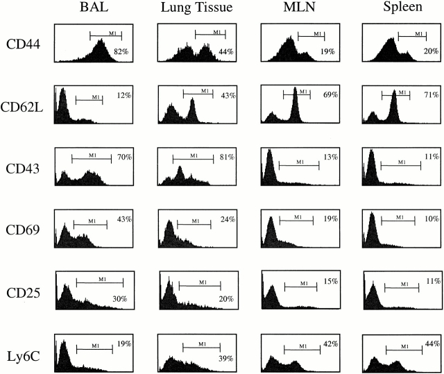
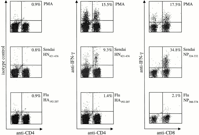
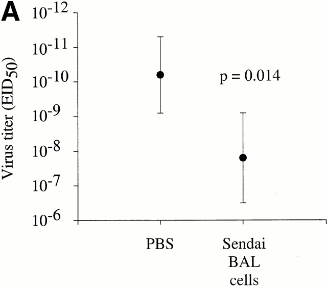
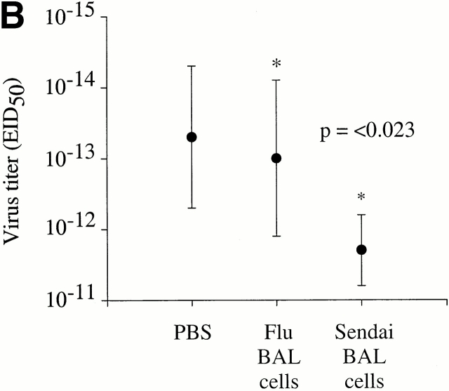
Although CD4(+) T cells have been shown to mediate protective cellular immunity against respiratory virus infections, the underlying mechanisms are poorly understood. For example, although phenotypically distinct populations of memory CD4(+) T cells have been identified in different secondary lymphoid tissues, it is not known which subpopulations mediate protective cellular immunity. In this report, we demonstrate that virus-specific CD4(+) T cells persist in the lung tissues and airways for several months after Sendai virus infection of C57BL/6 mice. A large proportion of these cells possess a highly activated phenotype (CD44(hi), CD62L(lo), CD43(hi), and CD25(hi)) and express immediate effector function as indicated by the production of interferon gamma after a 5-h restimulation in vitro. Furthermore, intratracheal adoptive transfer of lung memory cells into beta2m-deficient mice demonstrated that lung-resident virus-specific CD4(+) T cells mediated a substantial degree of protection against secondary virus infection. Taken together, these data demonstrate that activated memory CD4(+) T cells persisting at mucosal sites play a critical role in mediating protective cellular immunity.


Figures

Figure 1
Figure 1
CD4+ T cells in the lung airways (BAL) and lung tissues express an activated phenotype. Cells were isolated from the spleen, MLNs, lung tissue, and lung airways (BAL) of mice that had recovered from a Sendai virus infection (day 41 after infection). After isolation, the cells were stained with anti-CD4–PE and FITC-conjugated CD44, CD62L, CD43, CD69, and Ly6C antibodies. Cell surface expression of CD25 was determined using anti-CD25–PE and anti-CD4–FITC antibodies. The histograms show the expression of the indicated markers among live CD4+ lymphocytes. The bars and numbers in each panel show the percentage of CD4+ cells expres-sing the indicated marker. Data are representative of four independent experiments.
Figure 2
Figure 2
BAL memory cells produce IFN-γ directly ex vivo. Cells were isolated from the lung airways (BAL) of mice that had recovered from Sendai virus infection (day 34). The cells were restimulated with PMA/ionomycin or Sen-HN421–436, Flu-HA192–207, Sen-NP324–332, or Flu-NP366–374 peptides in the presence of BFA, IL-2, and naive Ly5.1+ spleen cells as APCs. After 5 h in culture, all cells were stained with anti-Ly5.2 biotin followed by streptavidin-allophycocyanin and the indicated antibodies. The data are presented as live Ly5.2+ lymphocytes (BAL). The numbers indicate the percentages of IFN-γ–secreting cells among the total Ly5.2+CD8+ or Ly5.2+CD4+ cell population. Data are representative of three independent experiments.
Figure 3
Figure 3
BAL memory cells confer protection against Sendai virus infection. (A) 10 naive C57BL/6 mice were intranasally infected with 500 EID50 Sendai virus. 4 h before infection, the mice received either memory BAL cells (5 × 10⁵ cells in 100 μl, n = 5), or control PBS (100 μl, n = 5) intratracheally. The BAL cells in this experiment were derived from C57BL/6 mice that had recovered from infection with 500 EID50 Sendai virus (35 d after infection). (B) 16 naive β2m-deficient mice were intranasally infected with 500 EID50 Sendai virus. 4 h before infection, each mouse received either Sendai memory BAL cells (106 cells in 100 μl, n = 6), influenza memory BAL cells (106 cells in 100 μl, n = 6), or control PBS (100 μl, n = 4) intratracheally. In this experiment, Sendai memory BAL cells were obtained from C57BL/6 mice at day 40 after infection, whereas influenza memory BAL cells were obtained from C57BL/6 mice at day 41 after infection. In both A and B, virus titers in the lung were determined 4 d after transfer by titrating lung homogenates in embryonated chicken eggs, followed by hemagglutination assays. Where indicated, the difference in viral titers among each group was determined to be statistically significant as based on a standard t test. The data are expressed as log10EID50 and are representative of three independent experiments.
Figure 3
Figure 3
BAL memory cells confer protection against Sendai virus infection. (A) 10 naive C57BL/6 mice were intranasally infected with 500 EID50 Sendai virus. 4 h before infection, the mice received either memory BAL cells (5 × 10⁵ cells in 100 μl, n = 5), or control PBS (100 μl, n = 5) intratracheally. The BAL cells in this experiment were derived from C57BL/6 mice that had recovered from infection with 500 EID50 Sendai virus (35 d after infection). (B) 16 naive β2m-deficient mice were intranasally infected with 500 EID50 Sendai virus. 4 h before infection, each mouse received either Sendai memory BAL cells (106 cells in 100 μl, n = 6), influenza memory BAL cells (106 cells in 100 μl, n = 6), or control PBS (100 μl, n = 4) intratracheally. In this experiment, Sendai memory BAL cells were obtained from C57BL/6 mice at day 40 after infection, whereas influenza memory BAL cells were obtained from C57BL/6 mice at day 41 after infection. In both A and B, virus titers in the lung were determined 4 d after transfer by titrating lung homogenates in embryonated chicken eggs, followed by hemagglutination assays. Where indicated, the difference in viral titers among each group was determined to be statistically significant as based on a standard t test. The data are expressed as log10EID50 and are representative of three independent experiments.

Comment in

References

    1. Doherty P.C., Topham D.J., Tripp R.A., Cardin R.D., Brooks J.W., Stevenson P.G. Effector CD4+ and CD8+ T-cell mechanisms in the control of respiratory virus infections. Immunol. Rev. 1997;159:105–117. - PubMed
    1. Kast W.M., Bronkhorst A.M., de Waal L.P., Melief C.J. Cooperation between cytotoxic and helper T lymphocytes in protection against lethal Sendai virus infection. Protection by T cells is MHC-restricted and MHC-regulated; a model for MHC-disease associations. J. Exp. Med. 1986;164:723–738. - PMC - PubMed
    1. Graham M.B., Braciale V.L., Braciale T.J. Influenza virus-specific CD4+ T helper type 2 T lymphocytes do not promote recovery from experimental virus infection. J. Exp. Med. 1994;180:1273–1282. - PMC - PubMed
    1. Graham M.B., Braciale T.J. Resistance to and recovery from lethal influenza virus infection in B lymphocyte–deficient mice. J. Exp. Med. 1997;186:2063–2068. - PMC - PubMed
    1. Zhong W., Marshall D., Coleclough C., Woodland D.L. CD4+ T cell priming accelerates the clearance of Sendai virus in mice, but has a negative effect on CD8+ T cell memory. J. Immunol. 2000;164:3274–3282. - PubMed

Publication types

MeSH terms

Substances