. 2001 Apr;107(8):995-1006.
doi: 10.1172/JCI11717.

Discordant effects of anti-VLA-4 treatment before and after onset of relapsing experimental autoimmune encephalomyelitis

Affiliations

Discordant effects of anti-VLA-4 treatment before and after onset of relapsing experimental autoimmune encephalomyelitis

B E Theien et al. J Clin Invest. 2001 Apr.

Abstract

Initial migration of encephalitogenic T cells to the central nervous system (CNS) in relapsing experimental autoimmune encephalomyelitis (R-EAE), an animal model of multiple sclerosis (MS), depends on the interaction of the alpha4 integrin (VLA-4) expressed on activated T cells with VCAM-1 expressed on activated cerebrovascular endothelial cells. Alternate homing mechanisms may be employed by infiltrating inflammatory cells after disease onset. We thus compared the ability of anti-VLA-4 to regulate proteolipid protein (PLP) 139-151-induced R-EAE when administered either before or after disease onset. Preclinical administration of anti-VLA-4 either to naive recipients of primed encephalitogenic T cells or to mice 1 week after peptide priming, i.e., before clinical disease onset, inhibited the onset and severity of clinical disease. In contrast, Ab treatment either at the peak of acute disease or during remission exacerbated disease relapses and increased the accumulation of CD4(+) T cells in the CNS. Most significantly, anti-VLA-4 treatment either before or during ongoing R-EAE enhanced Th1 responses to both the priming peptide and endogenous myelin epitopes released secondary to acute tissue damage. Collectively, these results suggest that treatment with anti-VLA-4 Ab has multiple effects on the immune system and may be problematic in treating established autoimmune diseases such as MS.


Figures
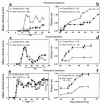
Figure 1
Figure 1
Anti–VLA-4 inhibits induction of, but exacerbates, ongoing clinical R-EAE. The indicated numbers of SJL mice were treated with rat control Ig or anti–VLA-4 (PS/2) beginning at day 7 (preclinical treatment), day 14 (acute-phase treatment), or day 24 (remission treatment) relative to priming with PLP139-151/CFA on day 0. Three treatments per week for 3 consecutive weeks were administered. (a and c) Short-term experiments from which animals were taken after three treatments and after nine treatments for immune assays (see Figures 4–6). Animals in experiments shown in b, d, e, and f also received nine treatments but were observed for a longer period of time. The data are expressed as mean clinical score versus day after priming (a, c, and e) and as the long-term relapse rate (b, d, and f). Data shown are representative of two to three separate experiments. AValues for the PS/2-treated mice are either significantly above or below those of the control Ig-treated mice; P < 0.05.
Figure 2
Figure 2
Anti–VLA-4 treatment of naive recipients provides short-term protection from expression of adoptive R-EAE. Ten recipient mice per group were treated with control Ig (days 1, 3, 5, 7, and 9) or with anti–VLA-4 relative to the transfer of 5 × 10⁶ PLP139-151–specific lymph node T-cell blasts on day 0. Anti–VLA-4 was administered on either days 1, 3, 5, 7, and 9 (a); days 3 and 5 (b); days 5 and 7 (c); or days 7 and 9 (d). AMean clinical scores of the PS/2-treated mice were significantly less than those of the control Ig-treated mice; P < 0.05.
Figure 3
Figure 3
Preclinical administration of anti–VLA-4 blocks infiltration of CD4+ T cells into the CNS. Lower thoracic spinal cord, cerebellar, and brainstem tissue sections from representative animals of the treatment groups described in Figure 1a were examined for infiltration of inflammatory cells on day 13 after immunization. Three treatments of control Ab or anti–VLA-4 had been given by this time point. DAPI, in blue, stains all cells; CD4+ cells are marked in red. (a) Spinal cord CD4+ infiltrate in a naive animal. (b) Spinal cord CD4+ infiltrate in a control Ig-treated animal. (c) Spinal cord CD4+ infiltrate in an anti–VLA-4–treated animal. All sections are 6 μm thick. ×100.
Figure 4
Figure 4
Preclinical administration of anti–VLA-4 does not inhibit activation of peripheral PLP139-151–specific T-cell responses. Splenic lymphocytes from three mice per group were harvested day 13 after immunization and tested for PLP139-151–specific proliferative and Th1 cytokine responses. Three treatments with either anti–VLA-4 or control Ab had been administered by this time point. (a) Viable cells (5 × 10⁵/well) were cultured with the indicated concentrations of PLP139-151 for 4 days. Cultures were pulsed with 3H-TdR 20 hours before harvest. Data are presented as Δ cpm (3H-TdR incorporation in cultures containing peptide antigen, 3H-TdR incorporation in cultures containing medium). Stimulation indices (3H-TdR incorporation in cultures containing peptide antigen, 3H-TdR incorporation in cultures containing medium) are indicated above each bar. 3H-TdR incorporation in cultures containing medium only were 2,849 ± 183 and 8,850 ± 1,227 for control and anti–VLA-4 groups, respectively. IL-2 (b) and IFN-γ (c) levels in the supernatants of cultures harvested at 24 and 48 hours after stimulation with 25 μM of PLP139-151 were determined by ELISA as described in Methods. (d) DTH responses to both the initiating PLP139-151 peptide and the relapse-associated PLP178-191 peptide were evaluated in 4–5 mice treated with either control Ig or anti–VLA-4 at day 36 after immunization. Data represent the mean 24-hour change in ear thickness ± SEM in response to challenge with 10 μg of each peptide. AIL-2 and IFN-γ levels of the PS/2-treated mice were significantly more than those of the control Ig-treated mice; P < 0.01.
Figure 5
Figure 5
Administration of anti–VLA-4 during ongoing R-EAE enhances proliferation and cytokine production to PLP139-151 and enhances epitope spreading to the relapse-associated PLP178-191 epitope. Spleen and lymph node cells from three mice per group were harvested at both 19 and 33 days after immunization from the experiment indicated in Figure 1c. (a) Viable lymph node cells (5 × 10⁵/well) from mice at day 19 after immunization (these mice had received three treatments with PS/2 beginning on day 14) were cultured with indicated concentrations of PLP139-151 for 4 days and proliferation was assessed by incorporation of 3H-TdR. 3H-TdR incorporation in cultures containing medium only were 1874 ± 286 and 1488 ± 334 for control and anti–VLA-4 groups, respectively. (b) Viable spleen cells (5 × 10⁵/well) from mice at day 33 after immunization (these mice had received nine treatments with PS/2 beginning on day 14) were cultured with indicated concentrations of PLP139-151 for 4 days and proliferation assessed by incorporation of 3H-TdR. 3H-TdR incorporation in cultures containing medium only was 4,199 ± 306 and 1,662 ± 248 for control and anti–VLA-4 groups, respectively. (c) Supernatants from the day 33 splenocyte cultures were harvested at 24 and 48 hours and analyzed for IFN-γ secretion by ELISA as described in Methods. Proliferative responses at day 33 after immunization to the relapse-associated PLP178-191 epitope were assessed from the lymph nodes (d) and the spleen (e). For d, 3H-TdR incorporation in cultures containing medium only was 2,214 ± 221 and 6,993 ± 1,274 for control and anti–VLA-4 groups, respectively. In e, medium-only 3H-TdR incorporation was 4,199 ± 306 and 1,662 ± 248 for control and anti–VLA-4 groups, respectively.
Figure 6
Figure 6
Mice treated with anti–VLA-4 during disease remission display increased autoantigen-specific Th1 responses to spread epitopes. DTH to PLP178-191, responses to which are associated with the primary relapse, and to MBP84-104, responses to which are associated with the second relapse peptide, were evaluated in mice treated with control Ig or anti–VLA-4 at day 40 (a) and/or day 66 (b) after immunization. Data represent mean ± SEM of the change in ear thickness 24 hours after ear challenge with 10 μg peptide. ADTH responses of the PS/2-treated mice were significantly greater than those of the control Ig-treated mice; P < 0.05.
Figure 7
Figure 7
Administration of anti–VLA-4 during ongoing R-EAE enhances infiltration of CD4+ T cells into the CNS and upregulates the expression of VCAM-1. Lower thoracic spinal cord tissue sections from representative animals of the treatment groups described in Figure 1c were examined for infiltration of inflammatory cells on day 33 after immunization. Nine treatments (initiated on day 14) of control Ab or anti–VLA-4 had been given by this time point. Both animals shown had a clinical score of 2. DAPI, in blue, stains all cells; CD4+ cells and VCAM-1 are marked in red. (a) Spinal cord CD4+ infiltrate in a control Ab–treated animal. (b) Spinal cord CD4+ infiltrate in an anti–VLA-4–treated animal. (c) Spinal cord VCAM-1 expression in a control Ab–treated animal. (d) Spinal cord VCAM-1 expression in an anti–VLA-4–treated animal. Tissue from a naive animal was tested simultaneously with all three Ab’s and revealed the presence of only DAPI+ (blue) cells. All sections are 6 μm thick. ×100.
Figure 8
Figure 8
Anti–VLA-4 can mediate T-cell costimulation in both cis and trans. Naive BALB/c splenic T cells (5 × 10⁴/well) were cultured in 96-well microtiter plates with 5-μm polystyrene sulfate-coated latex microspheres conjugated with the indicated Ab’s as detailed in Methods. Cultures were pulsed with 1 μCi of 3H-TdR at culture initiation and harvested 72 hours later. (a) The concentrations used for the mAbs were: anti-CD3, 0.5 μg/ml; anti-CD28, 20 μg/ml; and anti–VLA-4, 2 μg/ml. The Ab’s were mixed before conjugation to the polystyrene microspheres, and the Ab concentration in all tubes was normalized to a total of 40 μg/ml using hamster control Ig. (b) Anti-CD3 (0.5 μg/ml) and anti-CD28 (20 μg/ml) were conjugated to polystyrene beads. Separate beads were conjugated with the various indicated concentrations of anti–VLA-4. Naive BALB/c splenic T cells (5 × 10⁴/well) were cultured with a combination of the anti-CD3/CD28 + anti–VLA-4 beads in a 1:2 ratio. Hamster Ig–coated beads were added to anti-CD3 and anti-CD3/28 groups to normalize the total number of beads per well. Similar patterns of proliferation were observed in five separate experiments.

Similar articles

Cited by

References

    1. Brown AM, McFarlin DE. Relapsing experimental allergic encephalomyelitis in the SJL/J mouse. Lab Invest. 1981;45:278–284. - PubMed
    1. McRae BL, Kennedy MK, Tan LJ, Dal Canto MC, Miller SD. Induction of active and adoptive chronic-relapsing experimental autoimmune encephalomyelitis (EAE) using an encephalitogenic epitope of proteolipid protein. J Neuroimmunol. 1992;38:229–240. - PubMed
    1. Lehmann PV, Sercarz EE, Forsthuber T, Dayan CM, Gammon G. Determinant spreading and the dynamics of the autoimmune T-cell repertoire. Immunol Today. 1993;14:203–208. - PubMed
    1. McRae BL, Vanderlugt CL, Dal Canto MC, Miller SD. Functional evidence for epitope spreading in the relapsing pathology of experimental autoimmune encephalomyelitis. J Exp Med. 1995;182:75–85. - PMC - PubMed
    1. Vanderlugt CL, Karandikar NJ, Bluestone JA, Miller SD. The functional significance of epitope spreading and its regulation by costimulatory interactions. Immunol Rev. 1998;164:63–72. - PubMed

Publication types

MeSH terms