. 2001 Apr;107(8):1017-23.
doi: 10.1172/JCI10858.

Critical role for thyroid hormone receptor beta2 in the regulation of paraventricular thyrotropin-releasing hormone neurons

Affiliations

Critical role for thyroid hormone receptor beta2 in the regulation of paraventricular thyrotropin-releasing hormone neurons

E D Abel et al. J Clin Invest. 2001 Apr.

Abstract

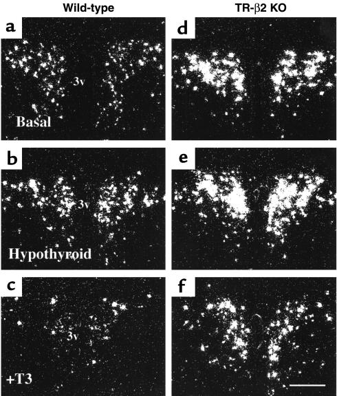
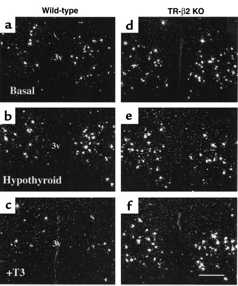
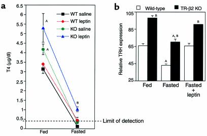
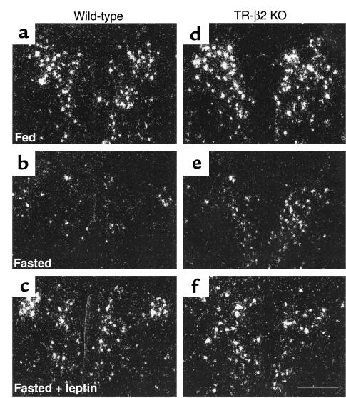
Thyroid hormone thyroxine (T(4)) and tri-iodothyronine (T(3)) production is regulated by feedback inhibition of thyrotropin (TSH) and thyrotropin-releasing hormone (TRH) synthesis in the pituitary and hypothalamus when T(3) binds to thyroid hormone receptors (TRs) interacting with the promoters of the genes for the TSH subunit and TRH. All of the TR isoforms likely participate in the negative regulation of TSH production in vivo, but the identity of the specific TR isoforms that negatively regulate TRH production are less clear. To clarify the role of the TR-beta2 isoform in the regulation of TRH gene expression in the hypothalamic paraventricular nucleus, we examined preprothyrotropin-releasing hormone (prepro-TRH) expression in mice lacking the TR-beta2 isoform under basal conditions, after the induction of hypothyroidism with propylthiouracil, and in response to T(3) administration. Prepro-TRH expression was increased in hypothyroid wild-type mice and markedly suppressed after T(3) administration. In contrast, basal TRH expression was increased in TR-beta2-null mice to levels seen in hypothyroid wild-type mice and did not change significantly in response to induction of hypothyroidism or T(3) treatment. However, the suppression of TRH mRNA expression in response to leptin reduction during fasting was preserved in TR-beta2-null mice. Thus TR-beta2 is the key TR isoform responsible for T(3)-mediated negative-feedback regulation by hypophysiotropic TRH neurons.


Figures

Figure 1
Figure 1
TSH response to PTU-induced hypothyroidism. Northern blot analysis of total RNA obtained from pooled pituitaries: n = 3 for wild-type (WT) and TR-β2–null (KO), demonstrating TSHβ mRNA responses in hypothyroid WT and KO mice. Amount of RNA loaded in each lane (in micrograms) is shown.
Figure 2
Figure 2
(af) Representative dark-field photomicrographs showing prepro-TRH mRNA in the rostral PVN of WT (ac) and TR-β2–null (KO) mice (df). Treatment conditions are as shown: basal (a and d), hypothyroid (b and e), and T3-treated (c and f). Scale bar, 300 μm.
Figure 3
Figure 3
(af) Representative dark-field photomicrographs showing prepro-TRH mRNA in the caudal PVN of WT (ac) and TR-β2–null (KO) mice (df). Treatment conditions are as shown: basal (a and d), hypothyroid (b and e), and T3-treated (c and f). Scale bar, 300 μm.
Figure 4
Figure 4
Relative prepro-TRH expression as assessed by laser densitometry in the rostral and caudal PVN of WT (open circles) and TR-β2–null (KO; filled circles) mice. Data shown are mean ± SEM. Each point represents analysis of anatomically identical sections from three separate animals. AP < 0.05 versus hypo and hyper WT, respectively, by ANOVA. BP < 0.05 versus WT in the same treatment group. Hypo, PTU–treated mice; Euth, untreated mice (basal conditions); Hyper, T3-treated mice.
Figure 5
Figure 5
Thyroid hormone and TRH responses to fasting and leptin administration. (a) Fed and fasting T4 concentrations in WT (black and red symbols) and TR-β2–null (KO) mice (green and blue symbols). n = 4 for all groups, except for saline-treated KO mice (n = 3). Data are mean ± SEM. AP < 0.05 versus fed WT; BP < 0.05 versus all other fasted mice by ANOVA. (b) Relative prepro-TRH expression as assessed by laser densitometry in the rostral PVN of WT (open bars) and TR-β2 KO mice (filled bars) at base line and after a 48- hour fast with and without leptin administration. Data shown are mean ± SEM. Each point represents analysis of anatomically identical sections. n = 3 fed groups, n = 3 fasted KO, n = 4 all other groups. AP < 0.05 versus fed or fasted plus leptin groups of same genotype, respectively. BP < 0.05 versus WT in the same treatment group by ANOVA.
Figure 6
Figure 6
Representative dark-field photomicrographs showing the response of prepro-TRH mRNA in the rostral PVN to the reduction in leptin levels during fasting in WT (ac) and KO mice (df). TRH mRNA decreased with fasting and was restored by leptin. Scale bar, 300 μm.

Similar articles

Cited by

References

    1. Larsen PR. Thyroid-pituitary interaction: feedback regulation of thyrotropin secretion by thyroid hormones. N Engl J Med. 1982;306:23–32. - PubMed
    1. Yamada M, et al. Tertiary hypothyroidism and hyperglycemia in mice with targeted disruption of the thyrotropin-releasing hormone gene. Proc Natl Acad Sci USA. 1997;94:10862–10867. - PMC - PubMed
    1. Taylor T, Weintraub BD. Altered thyrotropin (TSH) carbohydrate structures in hypothalamic hypothyroidism created by paraventricular nuclear lesions are corrected by in vivo TSH-releasing hormone administration. Endocrinology. 1989;125:2198–2203. - PubMed
    1. Chatterjee VK, Lee JK, Rentoumis A, Jameson JL. Negative regulation of the thyroid-stimulating hormone alpha gene by thyroid hormone: receptor interaction adjacent to the TATA box. Proc Natl Acad Sci USA. 1989;86:9114–9118. - PMC - PubMed
    1. Wondisford FE, et al. Thyroid hormone inhibition of human thyrotropin beta-subunit gene expression is mediated by a cis-acting element located in the first exon. J Biol Chem. 1989;264:14601–14604. - PubMed

Publication types

MeSH terms