. 2001 Apr 15;21(8):2669-77.
doi: 10.1523/JNEUROSCI.21-08-02669.2001.

Coexpression of microsomal-type prostaglandin E synthase with cyclooxygenase-2 in brain endothelial cells of rats during endotoxin-induced fever

Affiliations

Coexpression of microsomal-type prostaglandin E synthase with cyclooxygenase-2 in brain endothelial cells of rats during endotoxin-induced fever

K Yamagata et al. J Neurosci. .

Abstract

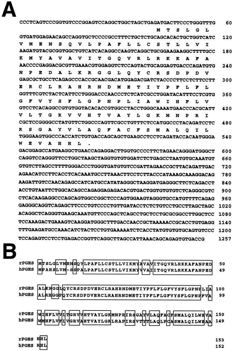
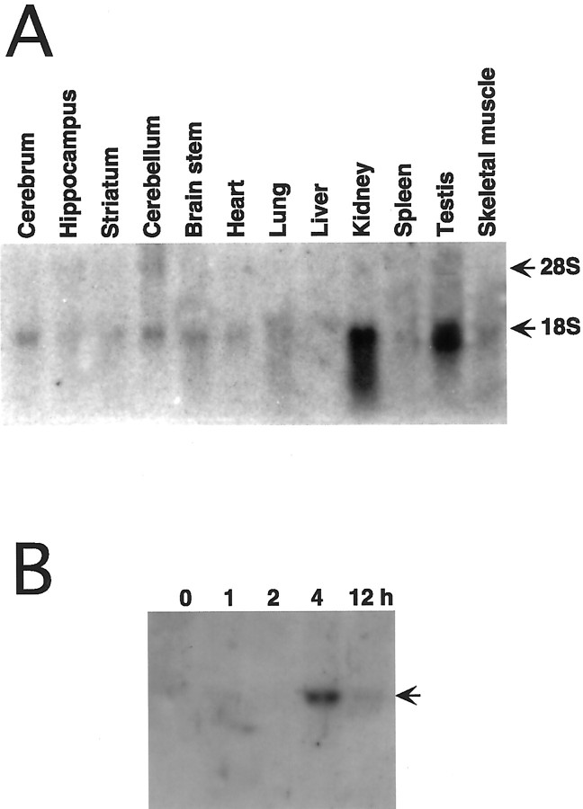
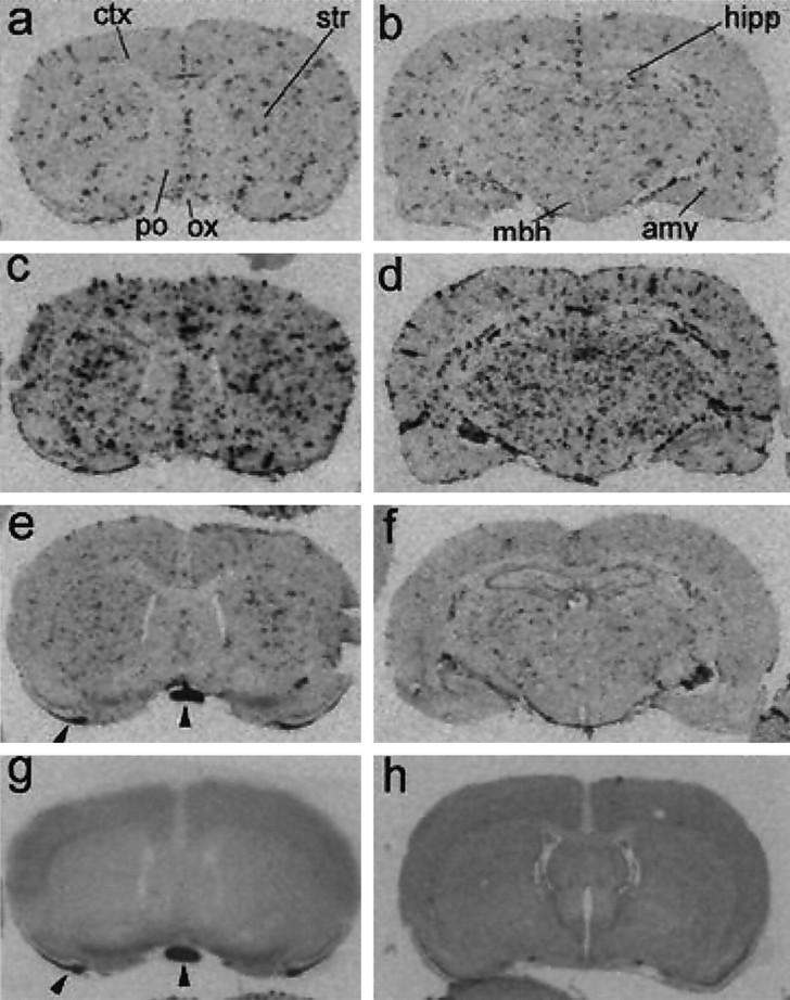
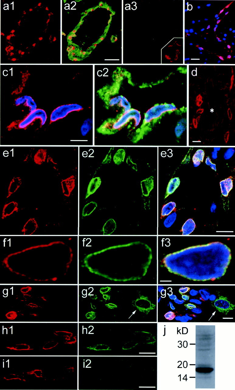
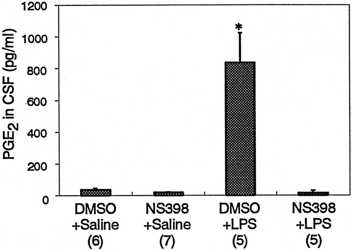
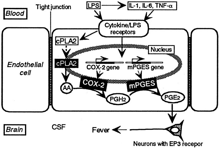
Fever is triggered by an elevation of prostaglandin E(2) (PGE(2)) in the brain. However, the mechanism of its elevation remains unanswered. We herein cloned the rat glutathione-dependent microsomal prostaglandin E synthase (mPGES), the terminal enzyme for PGE(2) biosynthesis, and examined its induction in the rat brain after intraperitoneal injection of pyrogen lipopolysaccharide (LPS). In Northern blot analysis, mPGES mRNA was weakly expressed in the brain under the normal conditions but was markedly induced between 2 and 4 hr after the LPS injection. In situ hybridization study revealed that LPS-induced mPGES mRNA signals were mainly associated with brain blood vessels, especially vein or venular-type ones, in the whole brain area. Immunohistochemical study demonstrated that mPGES-like immunoreactivity was expressed in the perinuclear region of brain endothelial cells, which were identified as von Willebrand factor-positive cells. Furthermore, in the perinuclear region of the endothelial cells, mPGES was colocalized with cyclooxygenase-2 (COX-2), which is the enzyme essential for the production of the mPGES substrate PGH(2). Inhibition of cyclooxygenase-2 activity resulted in suppression of both PGE(2) level in the CSF and fever (Cao et al., 1997), suggesting that the two enzymes were functionally linked and that this link is essential for fever. These results demonstrate that brain endothelial cells play an essential role in the PGE(2) production during fever by expressing COX-2 and mPGES.


Figures

Fig. 1.
Fig. 1.
A, Nucleotide sequence of ratmPGES cDNA and its predicted amino acid sequence. Numbering for nucleotides, on the right, is from the start site of the largest cDNA. Two independent cDNAs were sequenced on opposite strands and yielded identical sequences. B, Comparison of the deduced amino acid sequences of rat (rPGES) and human (hPGES) PGES. Conserved amino acids between rat and human PGES are boxed.
Fig. 2.
Fig. 2.
Expression and regulation of rat mPGESmRNA. Northern blot analysis of 30 μg of total RNA per lane prepared from brain and peripheral tissues. A, Tissue distribution analysis shows that mPGES mRNA expression is enriched in the testis and kidney, with a low level of expression detected in the brain regions. B, Forebrain RNA at different time points (0, 1, 2, 4, and 12 hr) after LPS injection (0.4 mg/kg, i.p.) reveal that LPS induced a transient increase inmPGES mRNA that peaked at 4 hr and persisted through 8 hr.
Fig. 3.
Fig. 3.
Macroautoradiographic images of mPGESmRNA signals in coronal sections of the rat brain. By 2 hr after LPS injection (a, b), the spot-likemPGES mRNA signals appeared in the brain parenchyma and subarachnoidal space. By 4 hr after the injection (c,d), the spot-like mPGES mRNA signals markedly increased in both number and intensity. At 12 hr after LPS injection (e, f), the mRNA signals were reduced in intensity but still existed. mPGES mRNA signals were not detectable in the brain section obtained 4 hr after saline injection (g). Hybridization with the sense cRNA probe did not show the spot-like signals in a brain section obtained 4 hr after LPS injection (h). The coronal planes in the left panels and h contained cerebral cortex (ctx), striatum (str), preoptic area (po), and optic chiasma (ox). Those in b, d, and fcontained more caudal brain regions, including hippocampus (hipp), amygdala (amy), and mediobasal hypothalamus (mbh). Arrowheads ine and g indicate nonspecific signals occasionally seen in the white matter at the edge of sections.
Fig. 4.
Fig. 4.
Light microscopic views of localization ofmPGES mRNA. a–d, mPGES mRNA signals in the rostral part of the preoptic area 4 hr after saline injection (a), and 2 (b), 4 (c), and 12 (d) hr after the LPS injection.3v, The third ventricle; ox, optic chiasma. Arrows in b and cindicate mPGES mRNA-positive cells associated with blood vessels. Most other mPGES mRNA-positive cells not denoted by arrows were also associated with blood vessels when examined under higher magnification. e–k,mPGES mRNA signals in brain parenchyma (eh, j), subarachnoidal space (i), and choroid plexus (k) 4 hr after the LPS injection, except for h, which was sampled at 2 hr after the injection. Note that the artery-like blood vessels (arrowheads in h and i) were devoid of the mRNA signals. j, Less intensemPGES mRNA signals (arrows) were occasionally seen in unidentified cells in the brain parenchyma along with intense mRNA signals associated with blood vessels (arrowheads).dg, Dentate gyrus of the hippocampus. k,mPGES mRNA signals were also expressed in the choroid plexus (arrows) and associated blood vessels (asterisk). Scale bar (in d):a–d, k, 100 μm; e–j, 50 μm.
Fig. 5.
Fig. 5.
Immunostaining of mPGES, von Willebrand factor, and COX-2 in the rat brain 5 hr after LPS injection. In allpanels, red and blueindicate mPGES-like immunoreactivity and nuclear DNA staining, respectively. a1, mPGES staining is seen along the vessel wall. a2, mPGES staining is overlaid with von Willebrand factor staining (green) in the same brain section as in a1. a3, Incubation with preabsorbed anti-mPGES eliminated the staining in the same blood vessel seen in the adjacent section. a3,Inset, Control experiment for the double-immunostaining of mPGES and von Willebrand factor. Incubation of mPGES-stained section with nonimmunized sheep IgG instead of anti-von Willebrand factor resulted in virtually no staining of endothelial cytosol.b, mPGES-positive cells are seen along an axially cut blood vessel. c1, Magnified view of mPGES-positive cells with nuclear DNA staining. c2, von Willebrand factor staining (green) is overlaid on mPGES and nuclear staining in c1. d, mPGES is negative in a large artery (asterisk) but is positive in small blood vessels nearby. mPGES staining (e1, f1,g1) is overlaid with COX-2 immunostaining (e2, f2, g2) and further with nuclear staining (e3, f3,g3). f1–f3, Perinuclear colocalization of mPGES and COX-2 is confirmed in enlarged views.g1–g3, mPGES and COX-2 are colocalized only in endothelial cells but not in cortical neurons (arrow), in which only COX-2 is expressed. h1, h2,i1, i2, Double-immunostaining of mPGES (h1, i1) and COX-2 (h2,i2) was performed using either the ordinary antibody mixture (h1, h2) or the mixture preabsorbed with COX-2 antigen peptide (i1,i2). Note that only COX-2 staining was eliminated after preabsorption with COX-2 antigen peptide. j, Western blot analysis of rat kidney revealed that the antibody used for immunohistochemistry properly recognized rat mPGES. Scale bars:a1–a3, 50 μm; b, 20 μm;c1, c2, 5 μm; d,e1–e3, g1–g3, h1,h2, i1, i2, 10 μm; andf1–f3, 2 μm.
Fig. 6.
Fig. 6.
PGE2 level in the CSF 5 hr LPS injection and effect of a COX-2-specific inhibitor.Numbers in the parentheses indicate the number of animals examined. *p < 0.0001
Fig. 7.
Fig. 7.
Our current hypothesis on the signaling cascade leading to fever in which brain endothelial cells play a pivotal role. See Discussion for the details. Dashed arrowindicates translocation of cPLA2. Other arrows indicate the flow of either molecules or signals

References

    1. Blalock JE. A molecular basis for bidirectional communication between the immune and neuroendocrine systems. Physiol Rev. 1989;69:1–32. - PubMed
    1. Blatteis CM, Sehic E. Fever: how may circulating pyrogens signal the brain? News Physiol Sci. 1997;12:1–9.
    1. Breder CD, Saper CB. Expression of inducible cyclooxygenase mRNA in the mouse brain after systemic administration of bacterial lipopolysaccharide. Brain Res. 1996;713:64–69. - PubMed
    1. Cao C, Matsumura K, Yamagata K, Watanabe Y. Induction by lipopolysaccharide of cyclooxygenase-2 mRNA in rat brain; its possible role in the febrile response. Brain Res. 1995;697:187–196. - PubMed
    1. Cao C, Matsumura K, Yamagata K, Watanabe Y. Endothelial cells of the rat brain vasculature express cyclooxygenase-2 mRNA in response to systemic interleukin-1β: a possible site of prostaglandin synthesis responsible for fever. Brain Res. 1996;733:263–272. - PubMed

Publication types

MeSH terms