Involvement of PACAP receptor in primary afferent fibre-evoked responses of ventral roots in the neonatal rat spinal cord
- PMID: 11309249
- PMCID: PMC1572720
- DOI: 10.1038/sj.bjp.0703980
Involvement of PACAP receptor in primary afferent fibre-evoked responses of ventral roots in the neonatal rat spinal cord
Abstract
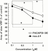
The role of PACAP receptor in nociceptive transmission was investigated in vitro using maxadilan, a PACAP receptor selective agonist and max.d.4, a PACAP receptor selective antagonist. Potentials, from a ventral root (L3 - L5) of an isolated spinal cord preparation or a spinal cord - saphenous nerve - skin preparation from 0 - 3-day-old rats, were recorded extracellularly. In the isolated spinal cord preparation, single shock stimulation of a dorsal root at C-fibre strength induced a slow depolarizing response lasting about 30 s (slow ventral root potential; slow VRP) in the ipsilateral ventral root of the same segment. Bath-application of max. d.4 (0.01 - 3 microM) inhibited the slow VRP in a concentration-dependent manner. In the spinal cord - saphenous nerve - skin preparation, application of capsaicin (0.1 microM) to the skin evoked a depolarization of the ventral root. This response was also depressed by max.d.4 (1 microM). Application of maxadilan evoked a long-lasting depolarization in a concentration-dependent manner in the spinal cord preparation. In the presence of max.d.4 (0.3 microM), the concentration response curve of maxadilan was shifted to the right. Reverse transcription-polymerase chain reaction (RT - PCR) experiments demonstrated the existence of PACAP receptor and VPAC(2) receptor in the neonatal rat spinal cord and [(125)I]-PACAP27 binding was displaced almost completely by maxadilan and max.d.4, but not by vasoactive intestinal peptide (VIP). These data indicate that PACAP receptor is dominantly distributed in the neonatal rat spinal cord. The present study suggests that PACAP receptor may play an excitatory role in nociceptive transmission in the neonatal rat spinal cord.
Figures


Similar articles
-
The excitatory and inhibitory modulation of primary afferent fibre-evoked responses of ventral roots in the neonatal rat spinal cord exerted by nitric oxide.Br J Pharmacol. 1996 Aug;118(7):1743-53. doi: 10.1111/j.1476-5381.1996.tb15600.x. Br J Pharmacol. 1996. PMID: 8842440 Free PMC article.
-
Muscarinic excitatory and inhibitory mechanisms involved in afferent fibre-evoked depolarization of motoneurones in the neonatal rat spinal cord.Br J Pharmacol. 1993 Sep;110(1):61-70. doi: 10.1111/j.1476-5381.1993.tb13772.x. Br J Pharmacol. 1993. PMID: 7693289 Free PMC article.
-
Effects of RP 67580, a tachykinin NK1 receptor antagonist, on a primary afferent-evoked response of ventral roots in the neonatal rat spinal cord.Br J Pharmacol. 1994 Dec;113(4):1141-6. doi: 10.1111/j.1476-5381.1994.tb17116.x. Br J Pharmacol. 1994. PMID: 7534180 Free PMC article.
-
Multiple receptors for PACAP and VIP.Trends Pharmacol Sci. 1994 Apr;15(4):97-9. doi: 10.1016/0165-6147(94)90042-6. Trends Pharmacol Sci. 1994. PMID: 7912462 Review. No abstract available.
-
Antidromic discharges of dorsal root afferents in the neonatal rat.J Physiol Paris. 1999 Sep-Oct;93(4):359-67. doi: 10.1016/s0928-4257(00)80063-7. J Physiol Paris. 1999. PMID: 10574124 Review.
Cited by
-
Effects of intrathecal administration of pituitary adenylate cyclase activating polypeptide on lower urinary tract functions in rats with intact or transected spinal cords.Exp Neurol. 2008 Jun;211(2):449-55. doi: 10.1016/j.expneurol.2008.02.022. Epub 2008 Mar 7. Exp Neurol. 2008. PMID: 18410926 Free PMC article.
-
Spinal astrocytic activation contributes to both induction and maintenance of pituitary adenylate cyclase-activating polypeptide type 1 receptor-induced long-lasting mechanical allodynia in mice.Mol Pain. 2016 May 12;12:1744806916646383. doi: 10.1177/1744806916646383. Print 2016. Mol Pain. 2016. PMID: 27175011 Free PMC article.
-
Spinal Astrocyte-Neuron Lactate Shuttle Contributes to the Pituitary Adenylate Cyclase-Activating Polypeptide/PAC1 Receptor-Induced Nociceptive Behaviors in Mice.Biomolecules. 2022 Dec 12;12(12):1859. doi: 10.3390/biom12121859. Biomolecules. 2022. PMID: 36551287 Free PMC article.
-
Analgesic effect of intrathecally administered orexin-A in the rat formalin test and in the rat hot plate test.Br J Pharmacol. 2002 Sep;137(2):170-6. doi: 10.1038/sj.bjp.0704851. Br J Pharmacol. 2002. PMID: 12208773 Free PMC article.
References
-
- ARIMURA A. Pituitary adenylate cyclase-activating polypeptide (PACAP): Discovery and current status of research. Reg. Peptides. 1992;37:287–303. - PubMed
-
- ARIMURA A. Receptors for pituitary adenylate cyclase activating polypeptide: comparison with vasoactive intestinal peptide receptors. Trends Endocrinol. Metab. 1993;3:288–294. - PubMed
-
- ARIMURA A. Perspectives on pituitary adenylate cyclase activating polypeptide (PACAP) in the neuroendocrine, endocrine, and nervous systems. Jpn. J. Physiol. 1998;48:301–331. - PubMed
-
- DICKINSON T., FLEETWOOD-WALKER S.M., MITCHELL R., LUTZ E.M. Evidence for roles of vasoactive intestinal polypeptide (VIP) and pituitary adenylate cyclase activating polypeptide (PACAP) receptors in modulating the responses of rat dorsal horn neurons to sensory inputs. Neuropeptides. 1997;31:175–185. - PubMed
-
- DICKINSON T., MITCHELL R., ROBBERECHT P., FLEETWOOD-WALKER S.M. The role of VIP/PACAP receptor subtypes in spinal somatosensory processing in rats with an experimental peripheral mononeuropathy. Neuropharmacology. 1999;38:167–180. - PubMed
MeSH terms
Substances
LinkOut - more resources
Full Text Sources

