. 2001 Apr 16;153(2):295-305.
doi: 10.1083/jcb.153.2.295.

OSP/claudin-11 forms a complex with a novel member of the tetraspanin super family and beta1 integrin and regulates proliferation and migration of oligodendrocytes

Affiliations

OSP/claudin-11 forms a complex with a novel member of the tetraspanin super family and beta1 integrin and regulates proliferation and migration of oligodendrocytes

S K Tiwari-Woodruff et al. J Cell Biol. .

Abstract

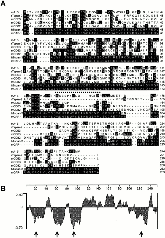
Oligodendrocyte-specific protein (OSP)/claudin-11 is a major component of central nervous system myelin and forms tight junctions (TJs) within myelin sheaths. TJs are essential for forming a paracellular barrier and have been implicated in the regulation of growth and differentiation via signal transduction pathways. We have identified an OSP/claudin-11-associated protein (OAP)1, using a yeast two-hybrid screen. OAP-1 is a novel member of the tetraspanin superfamily, and it is widely expressed in several cell types, including oligodendrocytes. OAP-1, OSP/claudin-11, and beta1 integrin form a complex as indicated by coimmunoprecipitation and confocal immunocytochemistry. Overexpression of OSP/claudin-11 or OAP-1 induced proliferation in an oligodendrocyte cell line. Anti-OAP-1, anti-OSP/claudin-11, and anti-beta1 integrin antibodies inhibited migration of primary oligodendrocytes, and migration was impaired in OSP/claudin-11-deficient primary oligodendrocytes. These data suggest a role for OSP/claudin-11, OAP-1, and beta1 integrin complex in regulating proliferation and migration of oligodendrocytes, a process essential for normal myelination and repair.


Figures

Figure 1
Figure 1
OAP-1 belongs to the tetraspanin family of proteins. (A) Multiple sequence alignment of OAP-1 with murine tetraspanins (mA15, mCD53, mCD63, mCD82), a rat tetraspanin (rTspan-2), and a human tetraspanin (hTspan-3). Boxed residues denote conservation among TM4SFs. (B) Kyte-Doolittle hydropathy plot of the predicted amino acid sequence of OAP-1, predicting four-transmembrane hydrophobic domains (arrows).
Figure 2
Figure 2
Presence of OAP-1 mRNA and protein in mouse brain and oligodendrocytes. (A) Northern blot analysis of OAP-1 expression. mRNA isolated from normal spinal cord (lane 1), brain (2), testes (3), skeletal muscle (4), heart (5), kidney (6), spleen (7), and lung (8) was probed with a 32P-labeled 1.9-kB cDNA of OAP-1. OAP-1 RNA was abundant in all tissue types. Gel loading was quantified with 18S expression. (B) Northern blot analysis of OAP-1 expression in mouse brain homogenate (BH) and purified oligodendrocytes (OL) from mouse brain. (C) Western blot analysis of OAP-1 protein in oligodendrocyte progenitor cells (O2A), purified mouse oligodendrocytes, and mouse brain homogenate. Preincubation of antibody with OAP-1–specific peptide eliminated the 31-kD band confirming antibody specificity (+Pep).
Figure 4
Figure 4
OAP-1 associates with OSP/claudin-11 and β1 integrin. (A) Mouse brain homogenates were immunoprecipitated with anti-OSP, anti–OAP-1, anti-GFAP, or anti–β1 integrin and then probed with anti–OAP-1 antibody. (B) Primary oligodendrocyte homogenates were immunoprecipitated with anti-OSP, anti–OAP-1, anti-GFAP, or anti–β1 integrin and then probed with anti–OAP-1 antibody. (C) Mouse brain homogenates were immunoprecipitated with anti-OSP, anti–OAP-1, anti-GFAP, or anti–β1 integrin and then probed with anti–OSP/claudin-11 antibody. (D) Mouse brain homogenates were immunoprecipitated with anti-OSP, anti-OAP-1, anti-GFAP, or anti–β1 integrin and then probed with anti–β1 integrin antibody. (E) CIMO homogenates were immunoprecipitated with anti-OSP, anti–β1 integrin, or anti-GFAP and then probed with anti–OAP-1 antibody. (F) Silver-stained gel of anti–OSP/claudin-11 (lane 1), anti–OAP-1 (lane 2), and anti–β1 integrin (lane 3) immunoprecipitated mouse brain homogenate. Small arrows denote bands that correspond to the molecular masses of OAP-1, OSP/claudin-11, and β1 integrin. The IgG band is visible at ∼50 kD.
Figure 3
Figure 3
In situ hybridization and immunohistochemistry showing OAP-1 expression in mouse forebrain. (A) Dark field photomicrograph of adult rat forebrain after in situ hybridization using 35S–OAP-1 cRNA shows dense neuronal expression of OAP-1 mRNAs in the CA1 region of a P10 mouse hippocampus. The area under the square is shown in B. (B) A high power bright field photomicrograph showing expression of OAP-1 mRNA in oligodendrocytes of the corpus callosum (arrowheads). (C) Using an immunohistochemistry anti–OSP/claudin-11 antibody (Texas red secondary antibody) showed staining primarily in white matter tracts and not in pyramidal cells and astrocytes (large arrow). Small arrows denote the outer borders of corpus callosum. (D) In contrast to OSP/claudin-11, immunohistochemistry using anti–OAP-1 antibody (FITC secondary antibody) in the same section demonstrated widespread staining in white matter tracts and in areas containing pyramidal cells and astrocytes (large arrow).
Figure 5
Figure 5
OSP/claudin-11 colocalizes with OAP-1 and β1 integrin in oligodendrocytes. (A) Confocal images of primary mouse oligodendrocytes shown in the top row were double labeled for OSP/claudin-11 (Texas red) and OAP-1 (FITC) or β1 integrin (FITC). Fused images are shown at the far right and bottom. Most cells showed striking colocalization of OSP/claudin-11, OAP-1, and β1 integrin. (B) Confocal images of primary mouse oligodendrocytes shown were double labeled for OAP-1 (Texas Red) and OSP/claudin-11 (FITC) in live primary oligodendrocytes. Fused images are showed to the far right. Colocalization of the two proteins was mostly observed in the cell borders. (C) Live primary oligodendrocytes were cultured on slides and immunostained with anti–OAP-1 (top row) and anti–OSP/claudin-11 (bottom row) antibody in the presence and absence of the respective antibody peptides. Background staining is also shown. Bars: (A, top row) 10 μM; (A, bottom row) 25 μM.
Figure 7
Figure 7
Overexpression of OSP/claudin-11 and OAP-1 increases proliferation in CIMO cells. (A) Northern blot of CIMO cells grown at 37°C (lanes 1 and 3) and 33°C (lanes 2 and 4) probed with a 32P-labeled 2-kb cDNA of OSP/claudin-11 (lanes 1 and 2) and 1.9-kB cDNA of OAP-1 (lanes 3 and 4). Both OSP/claudin-11 and OAP-1 expression is increased at 33°C during proliferation. (B) Western blot of CIMO cells transfected with control vector (lanes 1 and 3) and OSP (lanes 2 and 4) probed with anti-OSP antibody at 33°C (lanes 1 and 2) and 37°C (lanes 3 and 4). (Bottom panel) Western blot of CIMO cells transfected with OAP-1 (lane 1) and control vector (lane 2) at 37°C probed with anti–OAP-1 antibody confirms increased expression in these stable transfectents. (C) [3H]Thymidine incorporation into DNA of CIMO cells transfected with control vectors (pBabe and pcDNA) and OSP/claudin-11, antisense OSP/claudin-11, PLP, MBP, and OAP-1 constructs were observed under nonpermissive (37°C, minus Iγ) and permissive conditions (33°C, plus Iγ). OSP/claudin-11–transfected cells had a significant effect on proliferation at 33°C, whereas OAP-1 had a more generalized effect at both 33°C and 37°C. Control CIMO cells incorporated 4,000 ± 800 cpm/well/12 h, similar to the control vector transfected CIMO cells (average ± SEM, n = 24–32; 500 cells/well, each well counted in triplicates). OSP/claudin-11 versus pBabe vector at 37°C (***P < 0.0005), and OAP-1 versus pcDNA vector at 33°C and 37°C (**P < 0.005) (Student's t test).
Figure 6
Figure 6
OAP-1 and OSP/claudin-11 proteins are present on the cell surface. (A) Cell surface proteins of oligodendrocytes were biotinylated using a membrane-impermeant biotin ester. After solubilization and recovery of biotinylated proteins using Strepavidin-agarose, OAP-1 (lane 2) and OSP/claudin-11 (lane 3) proteins expressed at the plasma membrane were identified by SDS-PAGE and immunoblotting. Nonbiotinylated proteins (lane 1) were not observed. (B) Surface biotinylated oligodendrocyte proteins isolated from wild-type and OSP/claudin-11 transgenic mice were immunoprecipitated with anti–OAP-1 antibody (+/+, lane 1) and anti–OSP/claudin-11 antibody (+/+, lane 2; −/−, lane 3). Nonbiotinylated protein from wild-type oligodendrocytes was precipitated with anti–OSP/claudin-11 antibody (lane 4). Biotinylated proteins were visualized using Streptavidin-HRP as described in Materials and Methods. (C) Surface-biotinylated primary oligodendrocyte homogenates from wild-type and OSP/claudin-11 transgenic mice were immunoprecipitated with anti–OAP-1 antibody (+/+, lane 1) or anti–OSP/claudin-11 antibody (+/+, lane 2; −/−, lane 3) and probed with anti–OSP/claudin-11 antibody. (D) Surface-biotinylated primary oligodendrocyte homogenates from wild-type and OSP/claudin-11 transgenic mice were immunoprecipitated with anti–OAP-1 antibody (+/+, lane 1) or anti–OSP/claudin-11 antibody (+/+, lane 2; −/−, lane 3) and probed with anti–OAP-1 antibody. (E) Immunohistochemical EM of primary oligodendrocytes. OSP/claudin-11 and OAP-1 immunoreactivity was localized to the outer cell membrane (arrows). The asterisks represent the filter matrix the oligodendrocytes were grown on.
Figure 6
Figure 6
OAP-1 and OSP/claudin-11 proteins are present on the cell surface. (A) Cell surface proteins of oligodendrocytes were biotinylated using a membrane-impermeant biotin ester. After solubilization and recovery of biotinylated proteins using Strepavidin-agarose, OAP-1 (lane 2) and OSP/claudin-11 (lane 3) proteins expressed at the plasma membrane were identified by SDS-PAGE and immunoblotting. Nonbiotinylated proteins (lane 1) were not observed. (B) Surface biotinylated oligodendrocyte proteins isolated from wild-type and OSP/claudin-11 transgenic mice were immunoprecipitated with anti–OAP-1 antibody (+/+, lane 1) and anti–OSP/claudin-11 antibody (+/+, lane 2; −/−, lane 3). Nonbiotinylated protein from wild-type oligodendrocytes was precipitated with anti–OSP/claudin-11 antibody (lane 4). Biotinylated proteins were visualized using Streptavidin-HRP as described in Materials and Methods. (C) Surface-biotinylated primary oligodendrocyte homogenates from wild-type and OSP/claudin-11 transgenic mice were immunoprecipitated with anti–OAP-1 antibody (+/+, lane 1) or anti–OSP/claudin-11 antibody (+/+, lane 2; −/−, lane 3) and probed with anti–OSP/claudin-11 antibody. (D) Surface-biotinylated primary oligodendrocyte homogenates from wild-type and OSP/claudin-11 transgenic mice were immunoprecipitated with anti–OAP-1 antibody (+/+, lane 1) or anti–OSP/claudin-11 antibody (+/+, lane 2; −/−, lane 3) and probed with anti–OAP-1 antibody. (E) Immunohistochemical EM of primary oligodendrocytes. OSP/claudin-11 and OAP-1 immunoreactivity was localized to the outer cell membrane (arrows). The asterisks represent the filter matrix the oligodendrocytes were grown on.
Figure 8
Figure 8
Oligodendrocyte migration in the presence of OSP/claudin-11, OAP-1, and β1 integrin antibodies and in OSP/claudin-11–deficient cells. (A) Cells were migrated out of agarose drops in medium: −control, minus fibronectin; +control, plus fibronectin; anti–β1 integrin antibody; anti–OSP/claudin-11 antibody; anti–OAP-1 antibody; boiled inactive anti–OAP-1 antibody, I anti–OAP-1; anti-GFAP antibody; GRGDSP peptide; and GRGESP peptide. Except for the −control, all other conditions contained fibronectin. The total number of cells migrating out of the agar drop was counted after 4 d. Statistical values represent each condition versus positive control: **P < 0.0005; *P < 0.005 (Student's t test). (B) Migration assays were performed using oligodendrocytes isolated from wild-type (+/+), heterozygous OSP (+/−), and homozygous knockout OSP (−/−) mice, and cells were allowed to migrate in the presence (black bars) and absence (stippled bars) of fibronectin. There was marked attenuation of fibronectin-dependent migration in OSP/claudin-11–deficient cells. Values are expressed as percentage of wild-type. Heterozygous (+/−) versus wild-type (*P < 0.05) and homozygous recessive (−/−) versus wild-type (**P < 0.005) (Student's t test).

References

    1. Berditchevski F., Zutter M.M., Hemler M.E. Characterization of novel complexes on the cell surface between integrins and proteins with 4 transmembrane domains (TM4 proteins) Mol. Biol. Cell. 1996;7:193–207. - PMC - PubMed
    1. Blaschuk K.L., Frost E.E., Ffrench-Constant C. The regulation of proliferation and differentiation in oligodendrocyte progenitor cells by alphaV integrins. Development. 2000;127:1961–1969. - PubMed
    1. Bronner-Fraser M. Segregation of cell lineage in the neural crest. Curr. Opin. Genet. Dev. 1993;3:641–647. - PubMed
    1. Bronstein J.M., Popper P., Micevych P.E., Farber D.B. Isolation and characterization of a novel oligodendrocyte-specific protein. Neurology. 1996;47:772–778. - PubMed
    1. Bronstein J.M., Micevych P.E., Chen K. Oligodendrocyte-specific protein (OSP) is a major component of CNS myelin. J. Neurosci. Res. 1997;50:713–720. - PubMed

Publication types

MeSH terms

Associated data