. 2001 Apr 16;153(2):351-66.
doi: 10.1083/jcb.153.2.351.

Mammalian Abp1, a signal-responsive F-actin-binding protein, links the actin cytoskeleton to endocytosis via the GTPase dynamin

Affiliations

Mammalian Abp1, a signal-responsive F-actin-binding protein, links the actin cytoskeleton to endocytosis via the GTPase dynamin

M M Kessels et al. J Cell Biol. .

Abstract

The actin cytoskeleton has been implicated in endocytosis, yet few molecular links to the endocytic machinery have been established. Here we show that the mammalian F-actin-binding protein Abp1 (SH3P7/HIP-55) can functionally link the actin cytoskeleton to dynamin, a GTPase that functions in endocytosis. Abp1 binds directly to dynamin in vitro through its SH3 domain. Coimmunoprecipitation and colocalization studies demonstrated the in vivo relevance of this interaction. In neurons, mammalian Abp1 and dynamin colocalized at actin-rich sites proximal to the cell body during synaptogenesis. In fibroblasts, mAbp1 appeared at dynamin-rich sites of endocytosis upon growth factor stimulation. To test whether Abp1 functions in endocytosis, we overexpressed several Abp1 constructs in Cos-7 cells and assayed receptor-mediated endocytosis. While overexpression of Abp1's actin-binding modules did not interfere with endocytosis, overexpression of the SH3 domain led to a potent block of transferrin uptake. This implicates the Abp1/dynamin interaction in endocytic function. The endocytosis block was rescued by cooverexpression of dynamin. Since the addition of the actin-binding modules of Abp1 to the SH3 domain construct also fully restored endocytosis, Abp1 may support endocytosis by combining its SH3 domain interactions with cytoskeletal functions in response to signaling cascades converging on this linker protein.


Figures

Figure 1
Figure 1
Identification of proteins interacting with the SH3 domain of Abp1. (a) Blot overlay analysis using a GST-fusion protein containing the COOH-terminal half of Abp1 onto different rat tissue homogenates identified several bands prominent in brain at 75/80, 100, 145, and 180 kD. (b) Blot overlay analysis using a GST-fusion protein of the SH3 domain revealed bands of similar molecular weights not only in brain but in part also in testis and lung. (c) Affinity purifications of proteins interacting with the SH3 domains of Abp1, endophilin I, and syndapin I. Equal amounts of fusion proteins and brain extracts (0.625 mg brain protein per pull down) were used. 12.45 μg of starting material (corresponding to 1/50) were loaded for comparison. (d) Immunoprecipitated dynamin (arrow) was detected by both antidynamin antibodies (left) and by the Abp1-SH3 domain (right). Left two lanes in each gel: IP buffer with 150 mM salt; right two lanes in each gel: IP buffer with 100 mM salt.
Figure 2
Figure 2
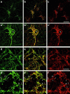
Colocalization of Abp1 and F-actin by confocal immunofluorescence microscopy. Primary hippocampal neurons kept in culture for 9 d displayed Abp1 accumulations in the extended peripheries of cell bodies as well as a cytosolic and neuritic immunostaining (a). F-actin was localized similarly (c), also seen in the merged image (b) and in the enlargements of the central areas of the images (a′–c′). (d–f) Neurons kept in culture for 20 d. At synapses, Abp1 (d) and F-actin (f) colocalize, as seen in the merged image (e) and in the enlargements of an area in the upper center of d–f (d′–f′). Stars in a–c and a′–c′ mark positions of cell bodies and arrows in d′–f′ mark selected examples of actin- and Abp1-rich postsynaptic structures. Bar: (c) 20 μm, (f) 10 μm.
Figure 5
Figure 5
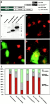
The Abp1 SH3 domain blocks endocytosis. (a) Schematic representation of myc-tagged Abp1 constructs used for transient transfections of Cos-7 cells. (b) SDS-PAGE and anti–myc immunoblotting of fusion proteins expressed in Cos-7 cells. The myc-SH3 construct (7 kD) is marked by a star. (c–e) Receptor-mediated endocytosis of Texas red-transferrin conjugates (red) in cells overexpressing Abp1 domains (green), merged images. (c) Untransfected cells. (d) Cells transfected with the Abp1-SH3 domain. (e) Cells overexpressing the actin-binding half of Abp1. Bar, 10 μm. (f) Quantitation of the results by assessing the percentages of cells lacking transferrin signal (block), displaying significantly reduced levels of uptake, and showing endocytosis capabilities similar to untransfected cells (data derived from group of cells showing high expression; data of lower expressing group of cells not shown, see text). Untransfected cells: 80.3 ± 6.0% normal, 12.7 ± 7.0 reduced, 7.3 ± 1.7% block, n = 359; myc-Grb2-SH3(N): 66.0 ± 8.0%, 23.5 ± 6.5%, 11.0 ± 1.0%, n = 359; myc-SH3: 16.5 ± 2%, 29.5 ± 0.5%, 54.5 ± 1.5%, n = 371; myc-flex/SH3mut: 53.0 ± 2.9%, 27.7 ± 3.3%, 19.3 ± 5.6%, n = 701; myc-ADF-H/hel: 70.8 ± 5.8%, 18.3 ± 4.7%, 11.3 ± 3.3%, n = 614; myc-ADF-H/hel/flex: 65.9 ± 10.0%, 17.9 ± 4.4%, 16.3 ± 5.9%, n = 587.
Figure 3
Figure 3
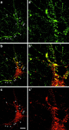
Colocalization of Abp1 (green) and dynamin (red) in primary hippocampal neurons by confocal microscopy. (a and a′) Abp1 strongly accumulated at defined sites in neurons kept in culture for 9 d (arrowheads). (c and c′) Besides cytosolic dynamin, sites of moderate dynamin accumulation proximal to the cell bodies were seen (arrowheads). These sites were immunopositive for Abp1, as seen in the merged images (b and b′). Note that subpools of both proteins did not spatially overlap (see neurites for Abp1 and cytoplasm for dynamin). a′–c′ are 2.5-fold enlargements of the center-right areas in a–c. Bar, 15 μm.
Figure 4
Figure 4
Coimmunoprecipitation of dynamin and Abp1. (a) Dynamin2aa-GFP coimmunoprecipitated (arrow) specifically with Flag-Abp1 from HEK cell extracts incubated with anti–Flag antibodies bound to protein G sepharose. Upper blots show material expressed by antitag immunostaining, middle blots show remaining protein in the post-IP supernatant, and bottom blots show immunoprecipitated Flag-Abp1 (left, arrowhead) and coimmunoprecipitated dynamin-GFP (right, arrow). Lysate and post-IP supernatant are 1/13 of the reaction. (b) Endogenous dynamin (detected by immunoblotting with the antidynamin antibodies) was specifically coimmunoprecipitated with Abp1 from rat brain extract with anti–Abp1 antibodies but not with equal amounts of unrelated guinea pig IgGs. Since Abp1 and IgGs both exhibit similar apparent molecular weights, the band of ∼55 kD represents both IgG and Abp1 (left lane), solely IgG (second from left lane), and, in the next two lanes, only Abp1 signals are observed at 55 kD. One third of immunoprecipitated material was loaded. Starting material represents 20 μg of brain protein.
Figure 6
Figure 6
Cooverexpression of dynamin rescues the endocytosis block caused by overexpression of the Abp1 SH3 domain. Cos-7 cells were double transfected with HA-dynamin1 and myc-Abp1 constructs. Quantification of transferrin uptake, as in Fig. 5 and Fig. 9, dark grey, no transferrin uptake, light grey, significantly reduced levels of uptake, and hatched cells show a transferrin uptake comparable with wild-type cells. Myc-(Abp1-)Flex/SH3–overexpressing cells were rescued up to a level similar to that caused by the flexible domain alone (a). Myc-(Abp1-)SH3–overexpressing cells were rescued up to a level similar to wild-type or HA-dynamin–overexpressing cells (b). Myc-flex/SH3: 19.5 ± 1.5% normal, 20.5 ± 1.5% reduced, 60.0 ± 3.0% block, n = 447; myc-flex/SH3 + HA-dynamin1: 68.7 ± 3.2%, 16.5 ± 2.5%, 14.75 ± 0.75%, n = 237; myc-flex/SH3mut: 53.0 ± 2.9%, 27.7 ± 3.3%, 19.3 ± 5.6%, n = 701; HA-dynamin1: 83.5 ± 3.7%, 13.7 ± 3.6%, 2.85 ± 0.05%, n = 213; hatched dark grey column: rescue effect, 81.0 ± 3.75 % (cotransfection rate of 96.5% was taken into consideration). This value represents 104.7 ± 8.7% of theoretically achievable maximal rescue (the fact that effect of SH3 domain but not that of the flexible domain can be rescued was taken into consideration; hatched light grey column). Myc-SH3: 16.5 ± 2%, 29.5 ± 0.5%, 54.5 ± 1.5%, n = 371; myc-SH3 + HA-dynamin1: 82.6 ± 2.3%, 12.7 ± 1.8%, 4.65 ± 0.45%; HA-dynamin1: 83.5 ± 3.7%, 13.7 ± 3.6%, 2.85 ± 0.05%, n = 213; hatched dark grey column: rescue effect (in case of myc-SH3 overexpression, identical to the achievable maximal rescue of SH3 domain effect), 96.5 ± 3.8% (cotransfection rate of 92.0% was taken into consideration).
Figure 7
Figure 7
Association of Abp1 with dynamin-containing pits upon growth factor stimulations of 3T3 fibroblasts. NIH3T3 fibroblasts were replated onto fibronectin-coated coverslips, stimulated with 300 ng/ml PMA and 5 ng/ml PDGF for 10 min, perforated with 0.02% saponin and subsequently fixed and processed for immunofluorescence microscopy (a–f). The lamellipodial accumulation of Abp1 (Kessels et al. 2000) was observable under these conditions (a and d). In addition, a widely distributed, punctate Abp1 immunostaining was detected (a and d). (c and f) Immunostaining of dynamin using the monoclonal antibody Hudy1 at perinuclear and cortical sites. (b and e) Merged images show that dynamin-containing sites are in almost all cases also immunopositive for Abp1 as especially well seen in extended lamellipodial areas (d–f). In contrast, in serum-starved cells, Abp1 (g) is readily extracted and the remaining protein shows no colocalization with dynamin (g–i). Inserts are enlargements of the marked areas. Bar, 10 μm.
Figure 9
Figure 9
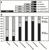
Sequential readdition of Abp1's actin-binding modules to the SH3 domain overexpression construct leads to a restoration of endocytosis in transfected Cos-7 cells. (a) Scheme of the overexpression constructs used. (b) Quantitation, myc-SH3: 16.5 ± 2% normal, 29.5 ± 0.5% reduced, 54.5 ± 1.5% block, n = 371; myc-flex/SH3: 19.5 ± 1.5%, 20.5 ± 1.5, 60.0 ± 1.5%, n = 447; myc-hel/flex/SH3: 56.7 ± 8.3%, 18.7 ± 4.9%, 24.3 ± 4.9%, n = 429; myc-Abp1(full length): 81 ± 7.8%, 13.0 ± 5.7%, 6.7 ± 1.7%, n = 448; untransfected cells: 80.3 ± 6.0%, 12.7 ± 7.0, 7.3 ± 1.7%, n = 359; myc-hel/flex/SH3mut: 70.3 ± 0.5%, 17.3 ± 1.3%, 11.7 ± 1.3%, n = 410.
Figure 8
Figure 8
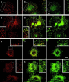
Cortical dynamin-rich sites in NIH3T3 cells, which become Abp1 immunopositive after receptor activation, are sites of endocytosis. Dynamin (a) and the endocytic coat component Hip1R (c) colocalize (b, merge) in cells stimulated with growth factors, as observed by confocal microscopy. Colocalization of dynamin (d) and eps15 (f), as seen in the merged image (e). Colocalization of AP2 (g) and Abp1 (i) at puncta at the cell cortex, but not at the leading edge of the cell; as seen in the merged image (h). (j–l) In nonperforated activated cells, the cytosolic pool of Abp1 largely obscures the weak Abp1 immunostaining (l) at sites of endocytosis [here marked by anti–AP2 immunostaining (j)]. Inserts represent enlargements of the areas boxed in a–l. Bars, 10 μm.
Figure 10
Figure 10
Schematic representation of Abp1 function at the interface of endocytosis and actin cytoskeleton.

References

    1. Ahn S., Maudsley S., Luttrell L.M., Lefkowitz R.J., Daaka Y. Src-mediated tyrosine phosphorylation of dynamin is required for beta2-adrenergic receptor internalization and mitogen-activated protein kinase signaling. J. Biol. Chem. 1999;274:1185–1188. - PubMed
    1. Brodin L., Löw P., Shupliakov O. Sequential steps in clathrin-mediated synaptic vesicle endocytosis. Curr. Opin. Neurobiol. 2000;10:312–320. - PubMed
    1. Cao H., Garcia F., McNiven M.A. Differential distribution of dynamin isoforms in mammalian cells. Mol. Biol. Cell. 1998;9:2595–2609. - PMC - PubMed
    1. Clark S.G., Stern M.J., Horvitz H.R. C. elegans cell-signaling gene sem-5 encodes a protein with SH2 and SH3 domains. Nature. 1992;356:340–344. - PubMed
    1. Damke H., Baba T., Warnock D.E., Schmid S.L. Induction of mutant dynamin specifically blocks endocytic coated vesicle formation. J. Cell Biol. 1994;127:915–934. - PMC - PubMed

Publication types

MeSH terms