. 2001 Apr 16;153(2):413-27.
doi: 10.1083/jcb.153.2.413.

Myopalladin, a novel 145-kilodalton sarcomeric protein with multiple roles in Z-disc and I-band protein assemblies

Affiliations

Myopalladin, a novel 145-kilodalton sarcomeric protein with multiple roles in Z-disc and I-band protein assemblies

M L Bang et al. J Cell Biol. .

Abstract

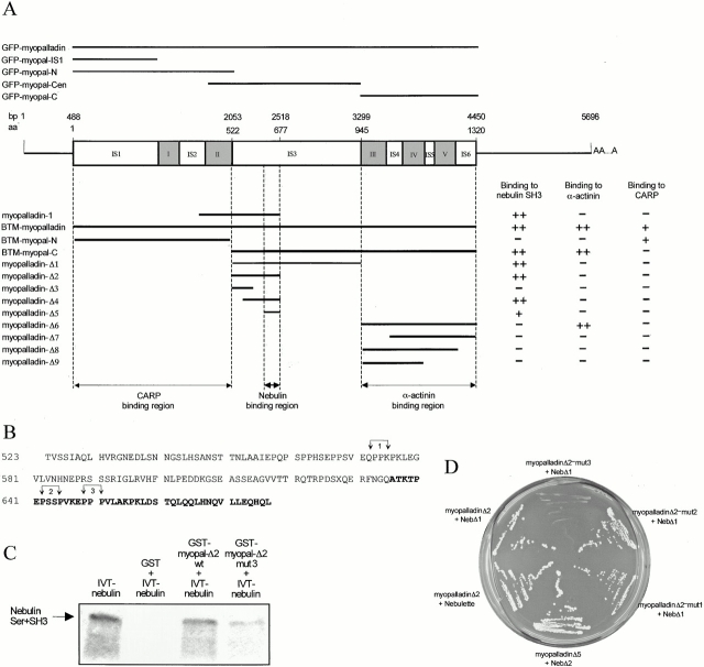
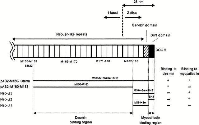
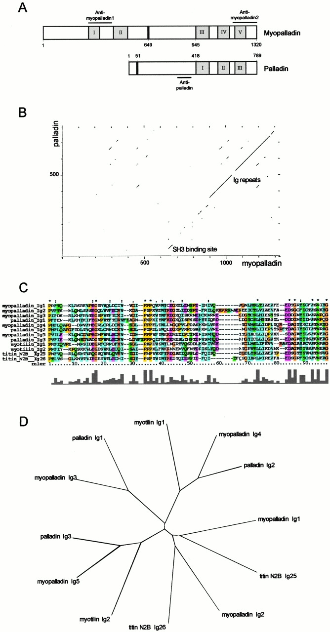
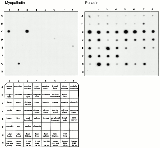
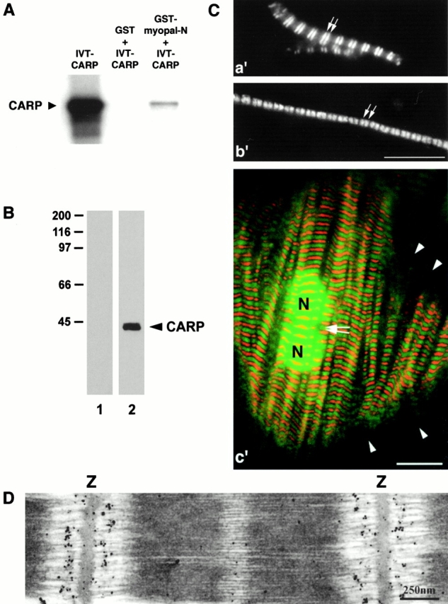
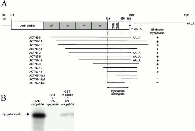
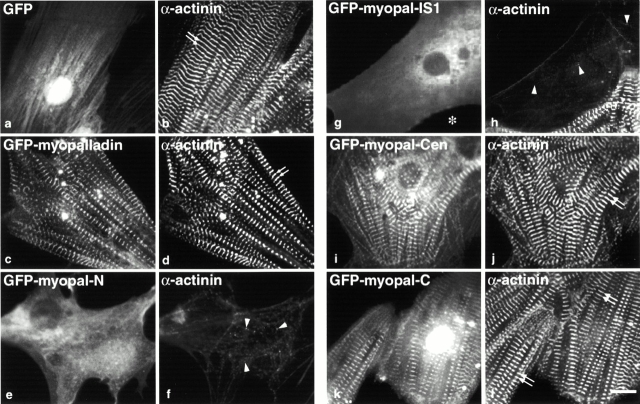
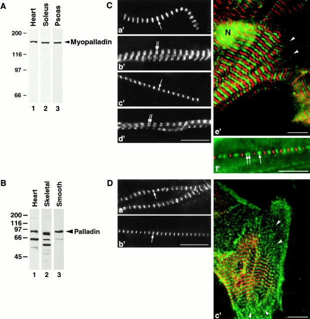
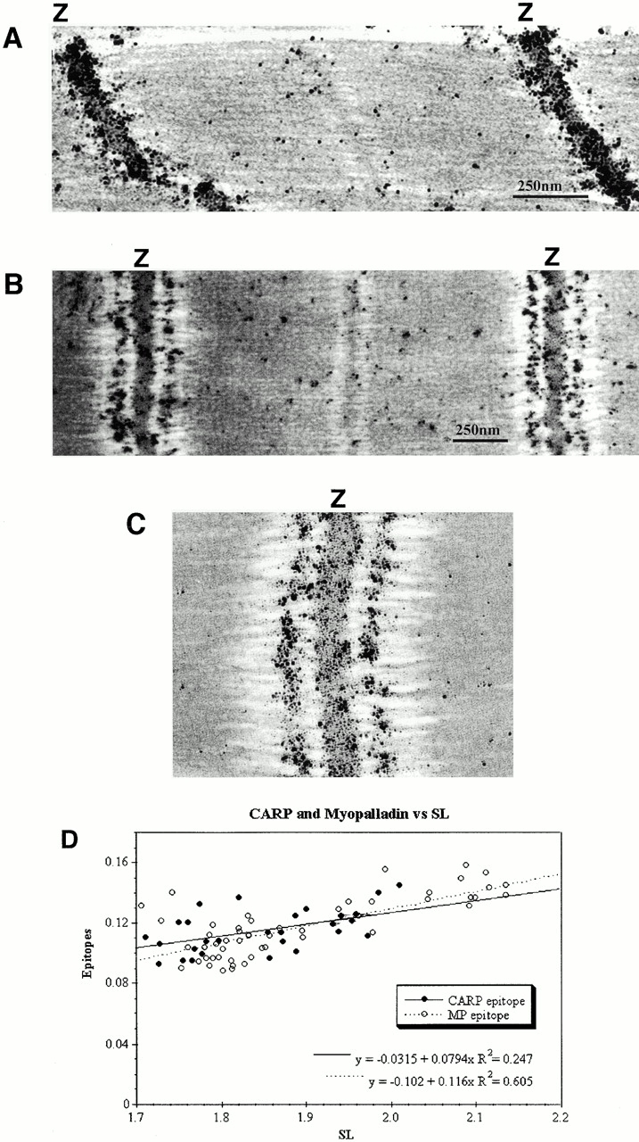
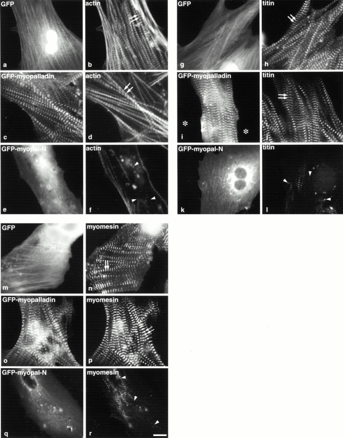
We describe here a novel sarcomeric 145-kD protein, myopalladin, which tethers together the COOH-terminal Src homology 3 domains of nebulin and nebulette with the EF hand motifs of alpha-actinin in vertebrate Z-lines. Myopalladin's nebulin/nebulette and alpha-actinin-binding sites are contained in two distinct regions within its COOH-terminal 90-kD domain. Both sites are highly homologous with those found in palladin, a protein described recently required for actin cytoskeletal assembly (Parast, M.M., and C.A. Otey. 2000. J. Cell Biol. 150:643-656). This suggests that palladin and myopalladin may have conserved roles in stress fiber and Z-line assembly. The NH(2)-terminal region of myopalladin specifically binds to the cardiac ankyrin repeat protein (CARP), a nuclear protein involved in control of muscle gene expression. Immunofluorescence and immunoelectron microscopy studies revealed that myopalladin also colocalized with CARP in the central I-band of striated muscle sarcomeres. Overexpression of myopalladin's NH(2)-terminal CARP-binding region in live cardiac myocytes resulted in severe disruption of all sarcomeric components studied, suggesting that the myopalladin-CARP complex in the central I-band may have an important regulatory role in maintaining sarcomeric integrity. Our data also suggest that myopalladin may link regulatory mechanisms involved in Z-line structure (via alpha-actinin and nebulin/nebulette) to those involved in muscle gene expression (via CARP).


Figures

Figure 4
Figure 4
Yeast two-hybrid screens reveal that myopalladin's nebulin-binding and α-actinin–binding sites are located in two distinct domains within its COOH-terminal 90-kD region. (A) Schematic structure of human full-length myopalladin cDNA. Numbers indicate nucleotide residue numbers (top, labeled bp) and amino acid residue numbers (below the nucleotide number, labeled aa). AAA indicates a poly(A+) tail. The Ig domains are shown in gray and are numbered I–V. Unique sequences are labeled IS1–6. Lines above the myopalladin domain structure indicate the myopalladin regions that were expressed as GFP fusion proteins in live cardiac myocytes (full-length myopalladin, two NH2-terminal fragments, one COOH-terminal fragment, and one internal fragment). Lines below indicate the partial myopalladin bait constructs that were generated for yeast two-hybrid screens to test for binding to the nebulin SH3 domain, α-actinin, and CARP. (+) and (−) denote the presence or absence of the growth of yeast colonies on SD/Trp-/Leu-/His-plates supplemented with 1.5 mM 3-AT. Myopalladin's nebulin-binding region was mapped to a part of IS3, whereas myopalladin's α-actinin–binding site was mapped to its COOH-terminal 375 residues. The CARP-binding site was assigned to myopalladin's NH2-terminal 522 residues. (B) The IS3 sequence of myopalladin contains three proline residue–rich motifs (labeled 1, 2, and 3). In each motif, pairs of proline residues (arrows) were mutated pairwise to glycine residues to obtain the bait constructs myopalladin-Δ2-mut1, 2, and 3. (C) Myopalladin interacted with nebulin in GST pull-down assays. In vitro–translated Ser+SH3 nebulin fragment (IVT-nebulin, lane 1) bound to glutathione-Sepharose 4B beads in the presence of GST-myopal-Δ2 wild-type (wt) fusion peptide (lane 3); a weaker binding was observed when proline residues 649 and 651 were mutated (lane 4; GST-myopal-Δ2-mut3 in B). As a negative control, binding of GST and nebulin Ser+SH3 to beads was tested (lane 2). (D) Interaction of myopalladin and nebulin/nebulette in the yeast two-hybrid system. Partial myopalladin cDNAs were cotransformed with partial nebulin/nebulette cDNAs into S. cerevisiae (see Fig. 3 A). The partial myopalladin sequence, myopalladin-Δ5 (IS3), was sufficient for binding to the SH3 domain of nebulin (Neb-Δ2). The myopalladin-Δ2-mut1,2 bait, but not the myopalladin-Δ2-mut3 bait interacted with nebulin preys, indicating that the PPP motif in myopalladin (residues 649–651) is involved in the interaction with nebulin. Additionally, myopalladin also bound to the SH3 domain of nebulette.
Figure 1
Figure 1
Yeast two-hybrid screens identify myopalladin and desmin as Z-line nebulin-binding proteins. Schematic structure of the COOH-terminal region of nebulin and its location within the sarcomere is shown based on previous immunoelectron microscopy studies (Millevoi et al. 1998). The nebulin COOH-terminal region constructs pAS2-M160-M185+Ser+SH3C and pAS2-M160-M183 identified myopalladin and desmin prey clones in yeast two-hybrid screens. Nebulin bait deletion constructs (NebΔ1–NebΔ3) allowed us to further map the myopalladin-binding region on nebulin to within the nebulin SH3 domain. The desmin-binding site on nebulin was found to be located within the nebulin M160-M183 modules. (+) and (−) denote the presence or absence of the growth of yeast colonies on SD/Trp-/Leu-/His-plates supplemented with 1.5 mM 3-AT.
Figure 2
Figure 2
Myopalladin is a novel 145-kD protein with homology to palladin. (A) Comparison of the domain architecture of myopalladin and the recently described palladin protein (Parast and Otey 2000). The Ig-I repeats are shown in gray, the unique sequences are shown in white, and the proline-rich PPP motif regions are shown in black. The bars above the myopalladin and below the palladin schematics indicate the recombinant fragments of the proteins that were used as antigens to generate specific antibodies (see Fig. 7 for the characterization of the antibodies). (B) Dot plot matrix sequence comparison of myopalladin and palladin. The regions of myopalladin and palladin that bind to the nebulin SH3 domain and to α-actinin are the most conserved regions between the two proteins. (C) Peptide sequence alignment of myopalladin's five Ig-I domains and comparison with Ig-I domains from myotilin (EMBL/GenBank/DDBJ accession number AF144477), titin N2B (accession number X90568), and palladin (accession number AB023209). All sequences are from humans. Identical residues are indicated by asterisks. (D) Phylogenetic tree based on the comparison of myopalladin's Ig-I repeats and the most related Ig-I repeats from myotilin, titin, and palladin.
Figure 3
Figure 3
Northern blot analysis reveals that detectable myopalladin gene expression is restricted to striated muscle. Myopalladin- and palladin-specific cDNA probes were hybridized to human RNA master blots (CLONTECH Laboratories, Inc.). Left, myopalladin gene expression is restricted to skeletal muscle (C3), fetal heart (G2), and adult heart (C1). Right, palladin transcripts are detected in all human tissues tested and are particularly abundant in striated and smooth muscle tissues (e.g., bladder, uterus, prostate, and stomach, C5–C8, respectively).
Figure 6
Figure 6
Identification of CARP as a sarcomeric component. (A) Interaction of myopalladin with CARP in GST pull-down assays. Full-length CARP was translated in vitro (left). When incubated together with GST-myopalladin fusion peptide (fragment myopal-N; see Fig. 4 A), binding to glutathione-Sepharose 4B beads was observed (right). Middle lane, negative control pull-down performed with GST and CARP peptides. (B) Specificity of affinity-purified anti-CARP antibodies. Western blot analyses of rat heart muscle lysates reveal that polyclonal antibodies raised to CARP react with a single 40-kD band (lane 2). No bands are detected in the control lane, which was incubated with secondary antibody alone (lane 1). (C) Immunofluorescence staining of CARP in washed isolated myofibrils from rat heart (a′) and skeletal (b′) muscle, as well as in primary cultures of rat cardiac myocytes (c′; CARP staining in green, myomesin staining in red), demonstrating that CARP is a sarcomeric component. Isolated myofibrils and cardiac myocytes were labeled with affinity-purified anti-CARP antibodies, followed by Cy2-conjugated secondary antibodies, and with antimyomesin antibodies (data not shown in a′ and b′) followed by Texas red–conjugated secondary antibodies. The merged images revealed that CARP is localized as a doublet within the I-band (in close proximity to the Z-line) in both heart and skeletal myofibrils and in isolated cardiac myocytes. Double arrows in a′ and b′ mark I-band staining. Arrowheads (in c′) indicate the absence of detectable CARP (and myomesin) staining in I-Z-I bodies, located at the edges of cultured cardiac myocytes. Note the additional staining of CARP in the nucleus in c′. N, nucleus. (D) Immunoelectron microscopy with CARP-specific antibodies demonstrates that CARP is present in the central I-band in cardiac myofibrils from mouse left ventricle. Z, Z-disc. Bars: (C) 10 μm; (D) 250 nm.
Figure 5
Figure 5
Myopalladin interacts with the EF hand region of α–actinin-2. (A) The schematic structure of α–actinin-2 (ACTN2) with its four central rod domains, R1–R4, and its unique terminal sequence is shown. Numbers indicate the nucleotide or amino acid residue numbers deduced from the human full-length cDNA sequence. AAA indicates a poly(A+) tail. A series of ACTN2 deletion constructs (below) were tested for interaction with full-length myopalladin in the yeast two-hybrid system. This assigned the myopalladin-binding domain within α-actinin to its COOH-terminal region containing the two EF hands. (+) and (−) denote the presence or absence of the growth of yeast colonies on SD-/Trp-/Leu-/His-plates supplemented with 1.5 mM 3-AT. (B) Interaction of myopalladin with α-actinin in GST pull-down assays. Myopalladin's COOH-terminal 376 residues were translated in vitro (IVT-myopal-Δ6, left). When incubated together with expressed ACTN14-GST fusion peptides, their binding to glutathione-Sepharose 4B beads was observed (right). Middle lane, negative control pull-down performed without the addition of the α-actinin fragment (for details, see Materials and Methods).
Figure 9
Figure 9
Overexpression of the NH2-terminal region of myopalladin in cardiac myocytes results in marked disruption of Z-line architecture as indicated by α-actinin staining. Cardiac myocytes expressing GFP alone (GFP; a), GFP–full-length myopalladin (GFP-myopalladin; c), GFP-myopalladin NH2-terminal region (GFP–myopal-N; e), a smaller GFP–NH2-terminal myopalladin IS1 region (GFP–myopal-IS1; g), GFP-myopalladin central region (GFP–myopal-Cen; i), and GFP-myopalladin COOH-terminal region (GFP–myopal-C; k) were fixed 3–5 d after transfection and stained with anti–α-actinin antibodies followed by Texas red–conjugated secondary antibodies (b, d, f, h, j, and l). Arrows point to the typical striated staining pattern of α-actinin in transfected cardiac myocytes (b, d, j, and l); arrowheads point to disrupted α-actinin staining pattern in transfected myocytes (f and h). Bar, 10 μm.
Figure 7
Figure 7
Characterization of endogenous myopalladins and palladins in striated muscle. (A) Specificity of affinity-purified antimyopalladin antibodies. Antimyopalladin antibodies recognize a band at ∼155 kD in rabbit heart (lane 1), soleus (lane 2), and psoas muscle (lane 3) by Western blot analysis. Note, on some blots an ∼35-kD band was also detected; the detection of this band was variable and its significance is unknown. (B) Specificity of affinity-purified antipalladin antibodies. Antipalladin antibodies recognize a 92-kD band in rat smooth muscle from the small intestine (lane 3), as well as multiple bands in heart (lane 1) and skeletal muscle (lane 2). (C) Immunofluorescence staining of myopalladin in washed, isolated myofibrils from rat heart (a′ and b′) and skeletal (c′ and d′) muscle, as well as in primary cultures of rat cardiac myocytes (e′; myopalladin staining in green, myomesin staining in red) demonstrating that myopalladin can be detected as a single striation at the Z-line (a′ and c′, arrows) and as a doublet within the I-band (in close proximity to the Z-line) (b′ and d′, double arrows). Isolated myofibrils and cardiac myocytes were labeled with affinity-purified antimyopalladin-1 antibodies, followed by Cy2-conjugated secondary antibodies, and with antimyomesin antibodies (data not shown in a′–d′) followed by Texas red–conjugated secondary antibodies. Note the additional staining of myopalladin in the nucleus in e′. Arrowheads in e′ mark the absence of detectable myopalladin (and myomesin) staining in I-Z-I bodies, located at the edges of cultured cardiac myocytes. (f′) Immunofluorescence image demonstrating the targeting of expressed GFP-myopalladin to the Z-line and to the I-band in primary cultures of chick cardiac myocytes. Cardiomyocytes expressing GFP–full-length myopalladin were fixed 3–5 d after transfection and stained with antimyomesin antibodies followed by Texas red–conjugated secondary antibodies and analyzed by immunofluorescence microscopy. Single arrows mark Z-line staining, whereas double arrows mark I-band staining. Significant variability in the relative labeling intensities of myopalladin at the Z-line vs. the I-band was observed. N, nucleus. (D) Immunofluorescence staining of palladin in washed, isolated myofibrils from rat heart (a′) and skeletal (b′) muscle, as well as in primary cultures of rat cardiac myocytes (c′; palladin staining in green, myomesin staining in red), demonstrating that palladin is localized at the Z-line. Isolated myofibrils and cardiac myocytes were labeled with affinity-purified antipalladin antibodies, followed by Cy2-conjugated secondary antibodies, and with antimyomesin antibodies (data not shown in a′ and b′) followed by Texas red–conjugated secondary antibodies. Arrowheads in c′ mark the presence of palladin (but not myomesin) staining in I-Z-I bodies, located at the edges of cultured cardiac myocytes. Note that palladin staining was not detected in the nucleus in c′. N, nucleus. Bars, 10 μm.
Figure 8
Figure 8
Immunoelectron microscopy reveals colocalization of myopalladin and CARP in mouse cardiac myofibrils. (A–C) Isolated myofibrils from mouse were prepared and stretched according to Trombitás et al. 1995; immunoelectron microscopy with affinity-purified antimyopalladin antibodies reveals that myopalladin is detected both within the periphery of the Z-line (A) and in the central I-band region (A–C). In stretched sarcomeres (B), the Z-line to I-band distance increases. A, unstretched; B, slightly stretched; C, high magnification view of Z-disc in stretched myofibrils. (D) The distances of I-band–bound CARP and myopalladin from the Z-disk were measured from immunoelectron micrographs at different sarcomere lengths (29 micrographs were analyzed for CARP; 54 micrographs for myopalladin). Both CARP and myopalladin colocalize in the central I-band and their epitope distances (in μm) from the Z-disc were dependent on sarcomeric length (SL).
Figure 10
Figure 10
Overexpression of the NH2-terminal region of myopalladin in cardiac myocytes results in disruption of thin, titin, and thick filaments. Chick cardiac myocytes expressing GFP alone (GFP; a, g, and m), GFP–full-length myopalladin (GFP-myopalladin; c, i, and o), and GFP–NH2-terminal myopalladin region (GFP–myopalladin-N; e, k and p) were fixed 3–5 d after transfection and stained with Texas red–conjugated phalloidin (b, d, and f), antititin T11 antibodies followed by Texas red–conjugated secondary antibodies (h, j, and l), or antimyomesin antibodies followed by Texas red–conjugated secondary antibodies (n, p, and r). Arrows point to typical striated staining pattern of actin (b and d), titin (h and j), and myomesin (n and p); arrowheads point to disrupted actin (f), titin (l), and myomesin (r). Bar, 10 μm.

References

    1. Arber S., Hunter J.J., Ross J., Jr., Hongo M., Sansig G., Borg J., Perriard J.C., Chien K.R., Caroni P. MLP-deficient mice exhibit a disruption of cardiac cytoarchitectural organization, dilated cardiomyopathy, and heart failure. Cell. 1997;88:393–403. - PubMed
    1. Baumeister A., Arber S., Caroni P. Accumulation of muscle ankyrin repeat protein transcript reveals local activation of primary myotube endcompartments during muscle morphogenesis. J. Cell Biol. 1997;139:1231–1242. - PMC - PubMed
    1. Beggs A.H., Byers T.J., Knoll J.H., Boyce F.M., Bruns G.A., Kunkel L.M. Cloning and characterization of two human skeletal muscle alpha-actinin genes located on chromosomes 1 and 11. J. Biol. Chem. 1992;267:9281–9288. - PubMed
    1. Centner T., Fougerousse F., Freiburg A., Witt C., Beckmann J.S., Granzier H., Trombitas K., Gregorio C.C., Labeit S. Molecular tools for the study of titin's differential expression. Adv. Exp. Med. Biol. 2000;481:35–49. - PubMed
    1. Chu W., Burns D.K., Swerlick R.A., Presky D.H. Identification and characterization of a novel cytokine-inducible nuclear protein from human endothelial cells. J. Biol. Chem. 1995;270:10236–10245. - PubMed

Publication types

MeSH terms

Associated data