. 2001 Apr 24;98(9):5276-81.
doi: 10.1073/pnas.091096198. Epub 2001 Apr 17.

Characterization of a high-voltage-activated IA current with a role in spike timing and locomotor pattern generation

Affiliations

Characterization of a high-voltage-activated IA current with a role in spike timing and locomotor pattern generation

D Hess et al. Proc Natl Acad Sci U S A. .

Abstract

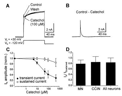
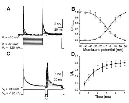
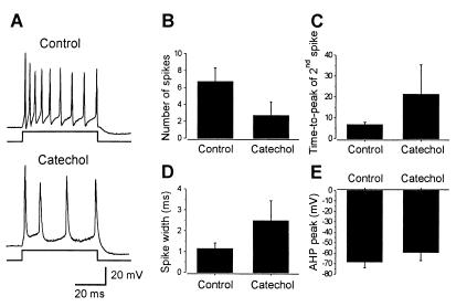
Transient A-type K+ channels (I(A)) in neurons have been implicated in the delay of the spike onset and the decrease in the firing frequency. Here we have characterized biophysically and pharmacologically an I(A) current in lamprey locomotor network neurons that is activated by suprathreshold depolarization and is specifically blocked by catechol at 100 microM. The biophysical properties of this current are similar to the mammalian Kv3.4 channel. The role of the I(A) current both in single neuron firing and in locomotor pattern generation was analyzed. The I(A) current facilitates Na+ channel recovery from inactivation and thus sustains repetitive firing. The role of the I(A) current in motor pattern generation was examined by applying catechol during fictive locomotion induced by N-methyl-d-aspartate. Blockade of this current increased the locomotor burst frequency and decreased the firing of motoneurons. Although an alternating motor pattern could still be generated, the cycle duration was less regular, with ventral roots bursts failing on some cycles. Our results thus provide insights into the contribution of a high-voltage-activated I(A) current to the regulation of firing properties and motor coordination in the lamprey spinal cord.


Figures

Figure 1
Figure 1
Pharmacology of the IA current. (A) Recordings from a MN in which catechol (100 μM) reversibly blocked the transient IA current without affecting the sustained current. (B) The subtracted current blocked by catechol. (C) Dose–response curves showing the effect of catechol on the IA and sustained currents. (D) Ratio of the transient to sustained current in MNs, CCINs, and all of the neurons studied.
Figure 2
Figure 2
Activation, inactivation, and recovery from inactivation of IA current. (A) A double test voltage step was applied in the control and in catechol (100 μM), the first to Vt between −60 and +30 mV and the second to +30 mV. IA current was isolated by subtracting the current evoked in catechol from that evoked in control. (B) The activation curve (■) was fitted with the best-fitting Boltzmann equation: G/Gmax = (1 + e(V − V1/2)/k)−1. The steady-state inactivation curve (○) was determined by plotting the mean normalized conductance as a function of the membrane potential. (C) A two-voltage-step protocol to +30 mV applied at increasing intervals was used to determine the recovery from inactivation of the IA current. The traces shown correspond to the current blocked by catechol. (D) The recovery from the inactivation curve corresponds to the mean normalized IA current amplitude as a function of the interpulse duration.
Figure 3
Figure 3
Role of IA current in repetitive firing in neurons in culture. (A) Application of a depolarizing current to isolated neurons in culture elicited repetitive firing of action potentials. Catechol blocked the repetitive firing. The membrane potential of the neuron was held at −80 mV. (B) Relationship between the current injected and the number of spikes evoked in the control and catechol (100 μM). (C) Catechol reversibly increased the amplitude of the first action potential and decreased that of the second action potential when it occurred. (D) The time to peak of the first action potentials was unaffected by catechol, whereas it was reversibly delayed for the second action potential. (E) The width of action potentials was reversibly increased by catechol. (F) The amplitude of the fAHP was reversibly reduced by catechol.
Figure 4
Figure 4
Role of IA current in repetitive firing in neurons in the isolated spinal cord. (A) Application of a depolarizing current to neurons recorded in the intact spinal cord elicited repetitive firing of action potentials. Catechol reduced the repetitive firing. The neuron had a resting membrane potential of −75 mV. (B) Catechol reduced the number of action potentials elicited by current injection and increased the time to peak of the second action potential (C) and the width of action potentials (D). (E) The amplitude of the fAHP was reduced by catechol.
Figure 5
Figure 5
IA current facilitates Na+ channel recovery from inactivation. (A) Action potentials were elicited in a neuron in culture by a conditioning pulse followed by a test pulse with decreasing time intervals. Catechol decreased the repetitive firing induced by both the conditioning and test pulse. The membrane potential of the neuron was held at −80 mV. (B) In the control, the amplitude of action potentials induced by the test pulse was unaffected by decreasing the interval between the conditioning and test pulse. Catechol significantly decreased the amplitude of action potentials when the test pulse was delivered at ≤4.5 ms after the conditioned pulse. (C) Catechol significantly increased the time to peak when the interval between the conditioning and test pulse was ≤4.5. (D) The membrane potential at the start of the test pulse was not significantly changed by a decrease in the interval between the two pulses in the control and catechol.
Figure 6
Figure 6
Blockade of IA current affected the locomotor pattern. (A) Intracellular recording was made from a MN (trough potential = −70 mV), and alternating ventral root bursts were recorded in ipsilateral (i-vr) and contralateral (c-vr) ventral roots. Catechol increased the frequency of the locomotor rhythm, which became irregular. (B) Catechol decreased the cycle duration and the burst proportion (C). (D) The left–right phase relationship was unchanged in catechol. (E) The number of action potentials fired by MNs during each locomotor cycle was reduced by catechol.

References

    1. Arshavsky Y I, Orlovsky G N, Panchin Y V, Roberts A, Soffe S R. Trends Neurosci. 1993;16:227–232. - PubMed
    1. Marder E, Calabrese R L. Physiol Rev. 1996;76:687–717. - PubMed
    1. Grillner S, Ekeberg Ö, El Manira A, Lansner A, Parker D, Tegnér J, Wallén P. Brain Res Rev. 1998;26:184–197. - PubMed
    1. Delgado-Lezama R, Hounsgaard J. Prog Brain Res. 1999;123:57–63. - PubMed
    1. El Manira A, Tegnér J, Grillner S. J Neurophysiol. 1994;72:1852–1861. - PubMed

Publication types

MeSH terms

LinkOut - more resources