. 2001 May 1;21(9):2992-9.
doi: 10.1523/JNEUROSCI.21-09-02992.2001.

Kainate receptors regulate unitary IPSCs elicited in pyramidal cells by fast-spiking interneurons in the neocortex

Affiliations

Kainate receptors regulate unitary IPSCs elicited in pyramidal cells by fast-spiking interneurons in the neocortex

A B Ali et al. J Neurosci. .

Abstract

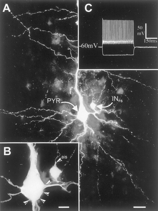
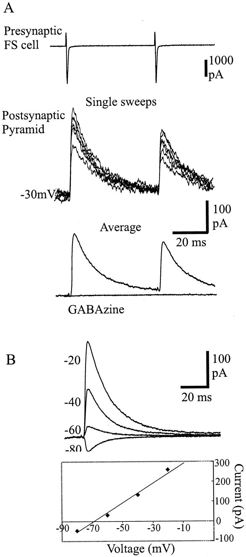
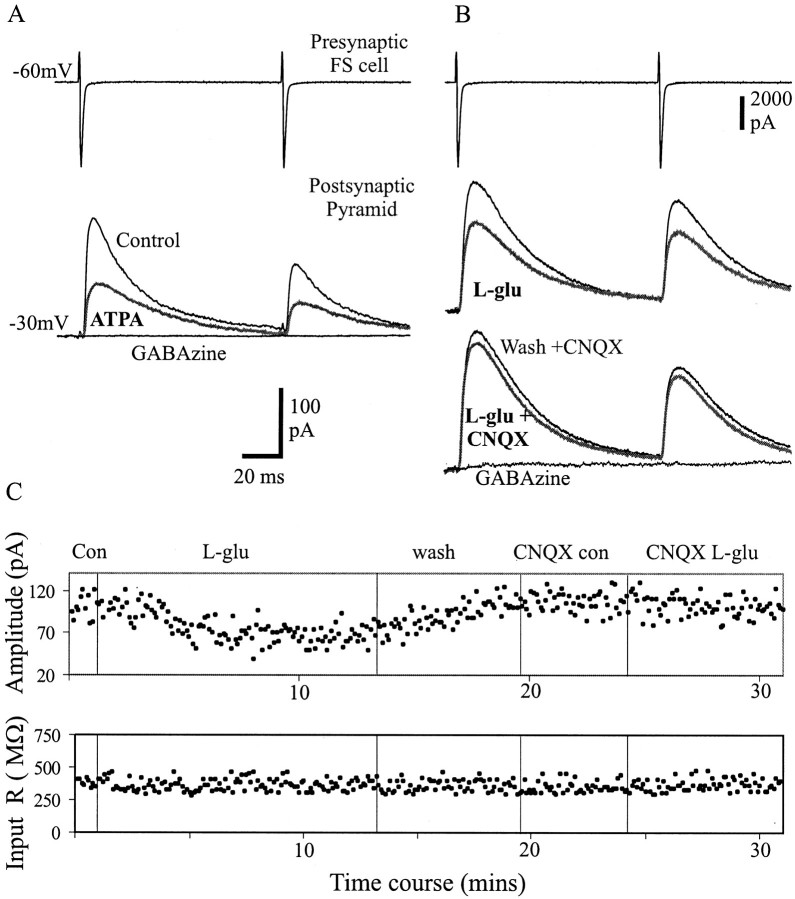
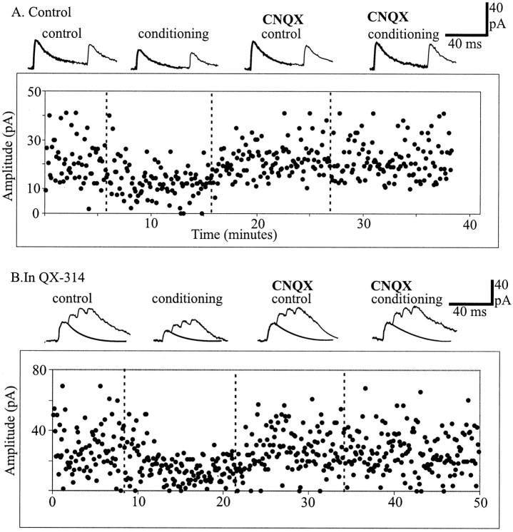
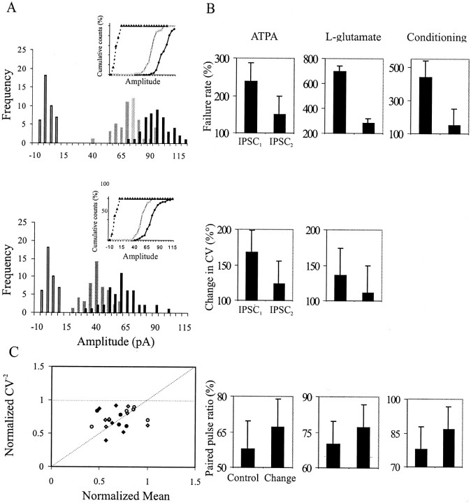
Unitary IPSCs elicited by fast-spiking (FS) interneurons in layer V pyramidal cells of the neocortex were studied by means of dual whole-cell recordings in acute slices. FS to pyramidal cell unitary IPSCs were depressed by (RS)-S-amino-3-(3-hydroxy-5-tert-butylisoxazol-4-yl) (ATPA), a kainate (KA) receptor agonist, and by the endogenous agonist l-glutamate in the presence of AMPA, NMDA, mGluR, and GABA(B) receptor antagonists. This effect was accompanied by an increase in failure rate of synaptic transmission, in the coefficient of variation, and in the paired pulse ratio, indicating a presynaptic origin of the IPSC depression. Pairing the activation of the presynaptic neuron with a depolarization of the postsynaptic cell mimicked the decrease of unitary IPSCs, and this effect persisted when postsynaptic sodium action potentials were blocked with the local anesthetic QX314. The effects of ATPA, glutamate, and of the pairing protocol were almost totally blocked by CNQX. These data suggest that KA receptors located on presynaptic FS cell terminals decrease the release of GABA and can be activated by glutamate released from the somatodendritic compartment of the postsynaptic pyramidal cells.


Figures

Fig. 1.
Fig. 1.
Extended-focus view reconstructions of a fast-spiking interneuron (INFS) and pyramidal cell (PYR) located in layer V of rat neocortex. A, Synaptically coupled FS interneuron to pyramidal cell. Note the small multipolar interneuron that gives rise to a dense axonal plexus (punctuate labeling) around the extremely large pyramidal cell. B, Higher magnification of the two cells. The axon initial segment (ais) of the interneuron is pointing toward the pial surface and emits a collateral, which is contacting the pyramidal cell four times (arrowheads), forming putative somatic synapses. This is an extended-focus view covering 50 μm in depth. C, Voltage responses of the presynaptic FS cell to −200 and +100 pA. The synaptic physiology of this pair is shown in Figure 2. Scale bars: A, 25 μm;B, 10 μm.
Fig. 2.
Fig. 2.
Characterization of a unitary connection between a fast-spiking cell and a pyramidal cell in layer V of the motor cortex.A, Top traces, Single sweep variation of IPSCs elicited in the pyramidal cell held at −30 mV by pairs of APs evoked in the FS cell. Bottom traces, Average IPSCs in control and in GABAzine (10 μm), a GABAA receptor antagonist. Note that the FS to pyramidal cell connections studied in layer V of the motor cortex typically displayed synaptic depression, which was frequency-dependent. B, Current–voltage relation of the unitary IPSCs characterized by a reversal potential of −70 mV.
Fig. 3.
Fig. 3.
Depression of unitary IPSCs by ATPA andl-glutamate. A, The selective kainate receptor agonist ATPA (1 μm) decreased the amplitude of the unitary IPSCs elicited in pyramidal cells (gray, bold trace). B, For another connection, the depression of unitary IPSCs was observed after application of the endogenous agonist l-glutamate (10 μm). All experiments were performed in the presence of GYKI 53655(50 μm) and d-AP-5 (50 μm), MCPG (1 mm), CPPG (100 μm), and CPG 55845 (100 μm) to block AMPA, NMDA, mGluR, and GABAB receptors, respectively. In B, naloxone (100 μm), atropine sulfate (50 μm), and DPCPX (1 μm) were also added to the drug cocktail to block opioid, muscarinic, and adenosine receptors, respectively. C,Plot of the peak amplitude of the first IPSCs and the input resistance of the postsynaptic cell for the connection shown in Bduring the time course of the experiment. Note that the input resistance did not significantly change during agonist application. Subsequent addition of CNQX (30 μm) almost completely abolished the suppression of the IPSCs induced byl-glutamate. Both connections in A andB were totally blocked by GABAzine.
Fig. 4.
Fig. 4.
Endogenous release of l-glutamate from the postsynaptic cell induced a depression of unitary IPSCs.A, B, Plot of the peak amplitude of the first IPSCs throughout experiments during which unitary synaptic responses were elicited in pyramidal cells alternatively in control conditions (control) and 900 msec after a conditioning depolarization of the postsynaptic pyramidal cells (conditioning). Insets illustrate the average unitary IPSCs elicited by pairs (A) or trains (B) of APs at different epochs of the experiments. Conditioning protocols with standard intracellular solution (A) or in the presence of QX-314 in the patch pipette (B) induced a decrease of the unitary IPSCs, which was prevented after subsequent addition of CNQX.
Fig. 5.
Fig. 5.
Origin of synaptic depression of unitary IPSCs. A, Example of the amplitude distribution and cumulative plot of first (top) and second (bottom) IPSCs in control (black) and during l-glutamate application (gray); open bars indicate noise. Single-sweep IPSC amplitudes are binned coarsely, and no peaks are distinguished. There is a clear shift in the mean amplitude after the application of l-glutamate. Billustrates the average change in failure rate and CV of first and second IPSCs and the PPR (IPSC2/IPSC1) before and during agonist application or during conditioning protocol. There was a greater increase in failure rate and CV for the first IPSCs compared with the second IPSCs. This resulted in an increase in PPR after agonist application and conditioning. C, To investigate the origin of the depression of IPSCs, a plot of normalized CV2(CV2 during KA receptor activation/control CV2) against the normalized mean, M (KA receptor activation IPSC/control IPSC), for the first (black symbols) and second (gray symbols) IPSC amplitude for 18 of the FS to pyramidal cell connections, assuming a simple binomial model. Each data point represents a pair in ATPA (diamonds) and l-glutamate (circles). A change of the amplitude without a change in the in CV2 would be caused by a change in q (quantal amplitude), represented by the zero slope dashed line. An equivalent change in the CV2 and M, represented by the line with the slope of 1, could be a change caused by n (number of release sites) and both the probability (p) and q. A greater change in CV-2 than in M would be attributable to a change in p with a slope >1. Most of the data points fall between these two lines (slope 0 and 1), indicating a change caused by a presynaptic origin, p.

References

    1. Alger BE, Pitler TA. Retrograde signalling at GABAA-receptor synapses in the mammalian CNS. Trends Neurosci. 1995;18:333–340. - PubMed
    1. Angulo MC, Rossier J, Audinat E. Postsynaptic glutamate receptors and integrative properties of fast-spiking interneurons in the rat neocortex. J Neurophysiol. 1999a;82:1295–1302. - PubMed
    1. Angulo MC, Staiger JF, Rossier J, Audinat E. Developmental synaptic changes increase the range of integrative capabilities of an identified excitatory neocortical connection. J Neurosci. 1999b;19:1566–1576. - PMC - PubMed
    1. Bahn S, Volk B, Wisden W. Kainate receptor gene expression in the developing rat brain. J Neurosci. 1994;14:5525–5547. - PMC - PubMed
    1. Bekkers JM, Richerson GB, Stevens CF. Origin of variability in quantal size in cultured hippocampal neurons and hippocampal slices. Proc Natl Acad Sci USA. 1990;87:5359–5362. - PMC - PubMed

Publication types

MeSH terms

LinkOut - more resources