. 2001 May 1;21(9):3017-23.
doi: 10.1523/JNEUROSCI.21-09-03017.2001.

Mitochondrial abnormalities in Alzheimer's disease

Affiliations

Mitochondrial abnormalities in Alzheimer's disease

K Hirai et al. J Neurosci. .

Abstract

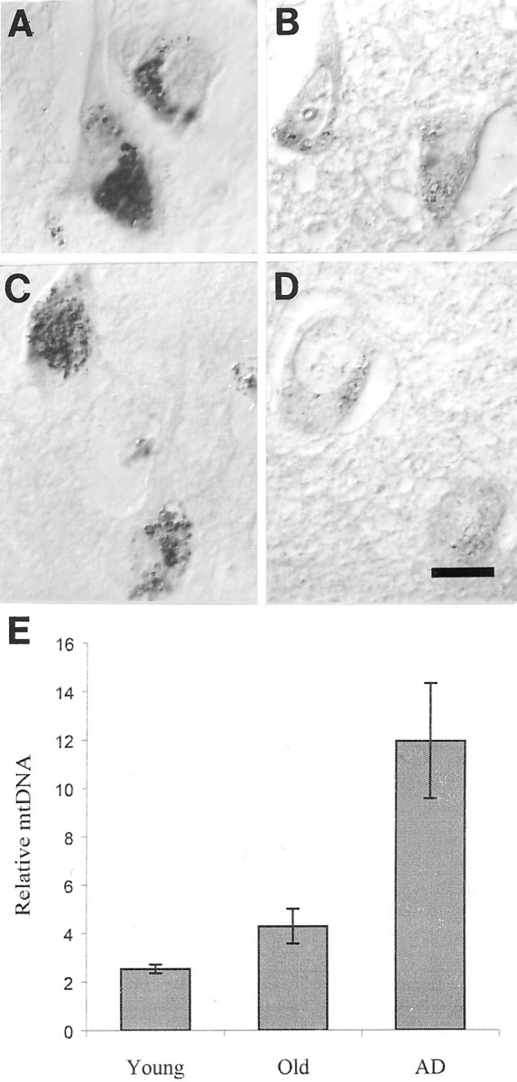
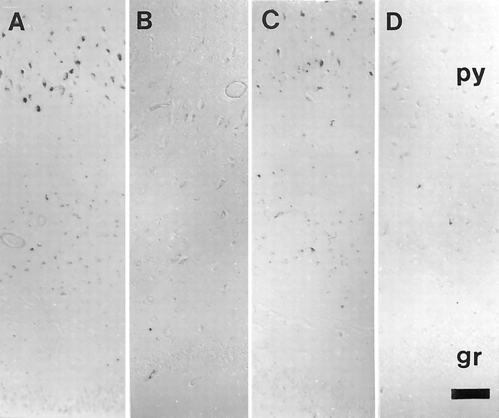
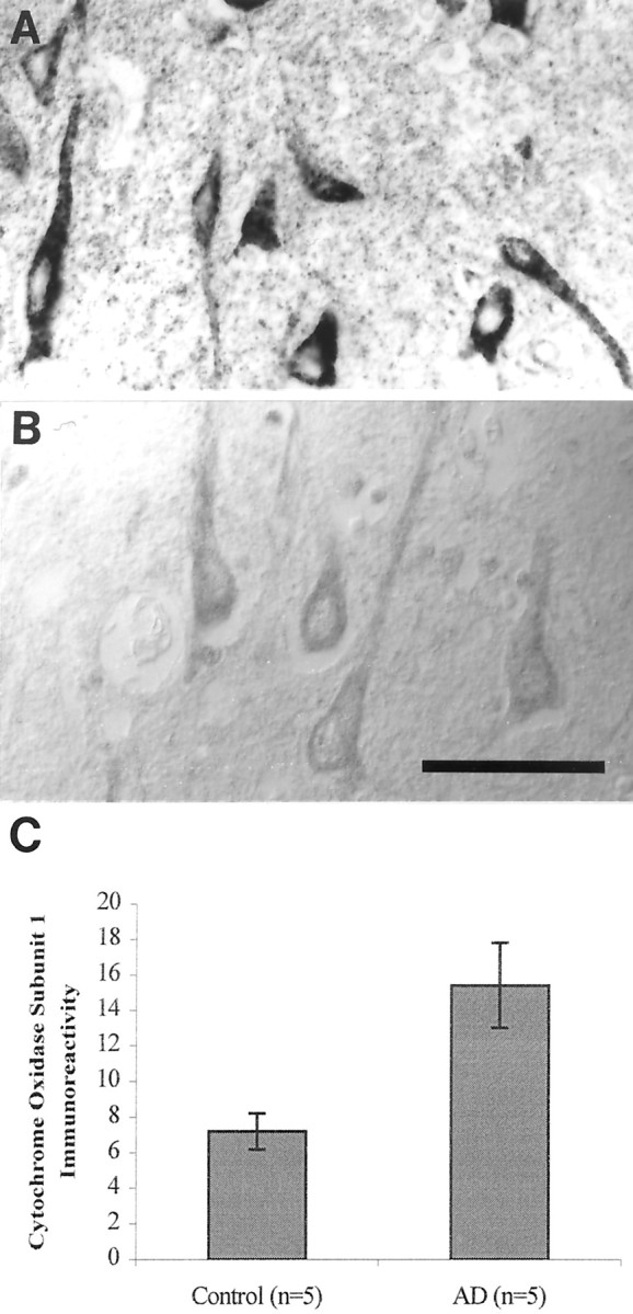
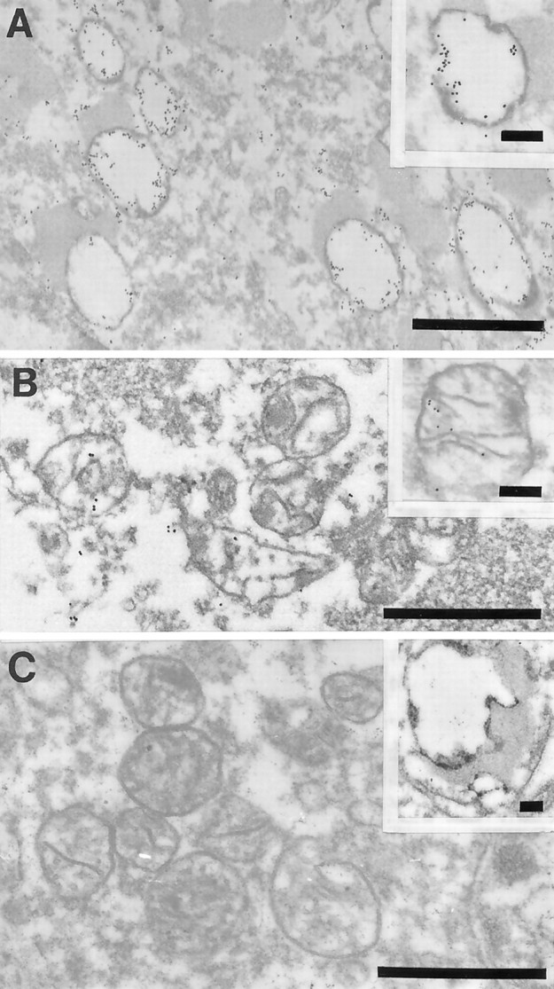
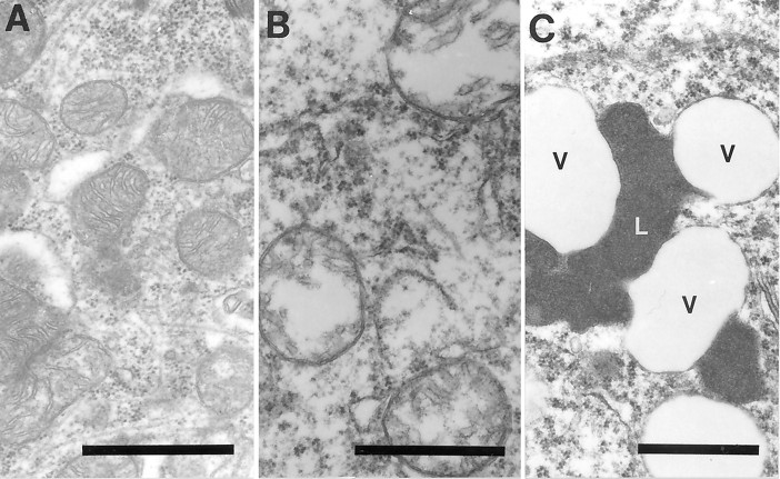
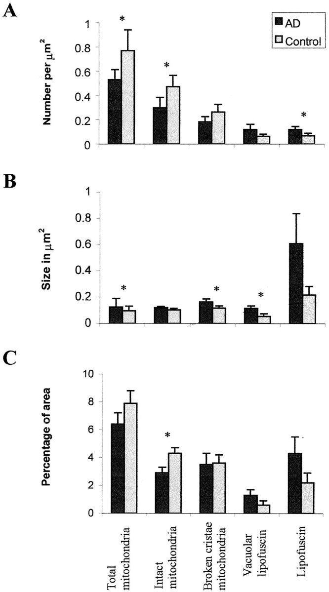
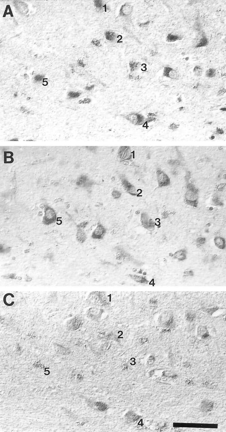
The finding that oxidative damage, including that to nucleic acids, in Alzheimer's disease is primarily limited to the cytoplasm of susceptible neuronal populations suggests that mitochondrial abnormalities might be part of the spectrum of chronic oxidative stress of Alzheimer's disease. In this study, we used in situ hybridization to mitochondrial DNA (mtDNA), immunocytochemistry of cytochrome oxidase, and morphometry of electron micrographs of biopsy specimens to determine whether there are mitochondrial abnormalities in Alzheimer's disease and their relationship to oxidative damage marked by 8-hydroxyguanosine and nitrotyrosine. We found that the same neurons showing increased oxidative damage in Alzheimer's disease have a striking and significant increase in mtDNA and cytochrome oxidase. Surprisingly, much of the mtDNA and cytochrome oxidase is found in the neuronal cytoplasm and in the case of mtDNA, the vacuoles associated with lipofuscin. Morphometric analysis showed that mitochondria are significantly reduced in Alzheimer's disease. The relationship shown here between the site and extent of mitochondrial abnormalities and oxidative damage suggests an intimate and early association between these features in Alzheimer's disease.


Figures

Fig. 1.
Fig. 1.
Pyramidal neurons of the hippocampus, cells highly vulnerable to death in AD, show increased mtDNA in all cases of AD (A, mtDNAΔ5kb, chimeric probe; C,wild-type mtDNA) compared with controls (B, mtDNAΔ5kb, chimeric probe; D, wild-type mtDNA). E,Quantitative densitometric analysis of the level of mtDNA shows the increases are severalfold and significant (p= 0.0034; Student's t test) for AD compared with old or young controls ± SEMs. Scale bar, 10 μm.
Fig. 2.
Fig. 2.
Both mtDNAΔ5kb (chimeric probe) (A, B) or wild-type mtDNA (wild type 1) (C, D) only show an increase in vulnerable neurons in AD. In the hippocampus, we found that, whereas pyramidal neurons (py) show mtDNA increase in AD (A, C) compared with controls (B, D), other neuronal populations, e.g., granule cells of the dentate gyrus (gr), as well as glia, show no detectable signals. Scale bar, 100 μm.
Fig. 3.
Fig. 3.
Cytochrome oxidase 1 immunoreactivity is increased severalfold in neurons in AD (A) compared with controls (B) and as shown by quantitative densitometric analysis (C) (p = 0.013; Student's ttest) ± SEM. Scale bar, 50 μm.
Fig. 4.
Fig. 4.
Ultrastructural examination of a pyramidal neuron in the hippocampus after in situ hybridization by using probes to wild mtDNA (wild type 1) in AD (A, B) or mtDNAΔ5kb (chimeric probe) (insets, A, B). The high density of gold particles was seen inside the vacuolar portions of lipofuscin granules, which likely represents autophagocytosis of damaged mitochondria in AD (A) and to a lesser extent in controls (C, inset). In contrast, mitochondria with cristae in both AD (B) and control cases (C) showed a lower level of mtDNA labeling (C). Scale bars: 1 μm; 0.25 μm (insets).
Fig. 5.
Fig. 5.
Examination of the morphology of mitochondria and lipofuscin in specimens removed at biopsy showed intact mitochondria (A), mitochondria with broken cristae (B), and vacuoles associates with lipofuscin indicated by a V and lipofuscin indicated by an L (C). Scale bars, 1 μm.
Fig. 6.
Fig. 6.
Morphometric analysis of the number (A), size (B), and percentage coverage (C) of cytoplasmic area by intact mitochondria, mitochondria with broken cristae, total mitochondria (intact plus mitochondria with broken cristae), vacuoles associated with lipofuscin, and lipofuscin in cases of AD and controls. Whereas percentage coverage of intact mitochondria decreases in AD (p = 0.012), no significant changes were noted in vacuoles associated with lipofuscin (p = 0.056), mitochondria with broken cristae (p > 0.10), or lipofuscin (p > 0.08). Although the size of lipofuscin did not change in AD (p = 0.095), that of the vacuoles associated with lipofuscin significantly increased (p = 0.029). Statistical comparison by theF test. * indicates significant difference between AD and control cases.
Fig. 7.
Fig. 7.
The distribution of neurons showing increased mtDNAΔ5kb (chimeric probe) (A), 8-OHG (B), and nitrotyrosine (C) immunoreactivity in AD completely overlaps. Numberindicates the same neurons in adjacent serial sections. Scale bar, 50 μm.

References

    1. Blass JP, Gibson GE. The role of oxidative abnormalities in the pathophysiology of Alzheimer's disease. Rev Neurol (Paris) 1991;147:513–525. - PubMed
    1. Blass JP, Baker AC, Ko L, Black RS. Induction of Alzheimer antigens by an uncoupler of oxidative phosphorylation. Arch Neurol. 1990;47:864–869. - PubMed
    1. Brunk UT, Jones CB, Sohal RS. A novel hypothesis of lipofuscinogenesis and cellular aging based on interactions between oxidative stress and autophagocytosis. Mutat Res. 1992;275:395–403. - PubMed
    1. Dowson JH, Mountjoy CQ, Cairns MR, Wilton-Cox H, Bondareff W. Lipopigment changes in Purkinje cells in Alzheimer's disease. J Alzheimer's Dis. 1998;1:71–79. - PubMed
    1. Friguet B, Stadtman ER, Szweda LI. Modification of glucose-6-phosphate dehydrogenase by 4-hydroxy-2-nonenal. Formation of cross-linked protein that inhibits the multicatalytic protease. J Biol Chem. 1994;269:21639–21643. - PubMed

Publication types

MeSH terms