. 2001 May 1;21(9):3104-12.
doi: 10.1523/JNEUROSCI.21-09-03104.2001.

Interleukin-10 prevents glutamate-mediated cerebellar granule cell death by blocking caspase-3-like activity

Affiliations

Interleukin-10 prevents glutamate-mediated cerebellar granule cell death by blocking caspase-3-like activity

A Bachis et al. J Neurosci. .

Abstract

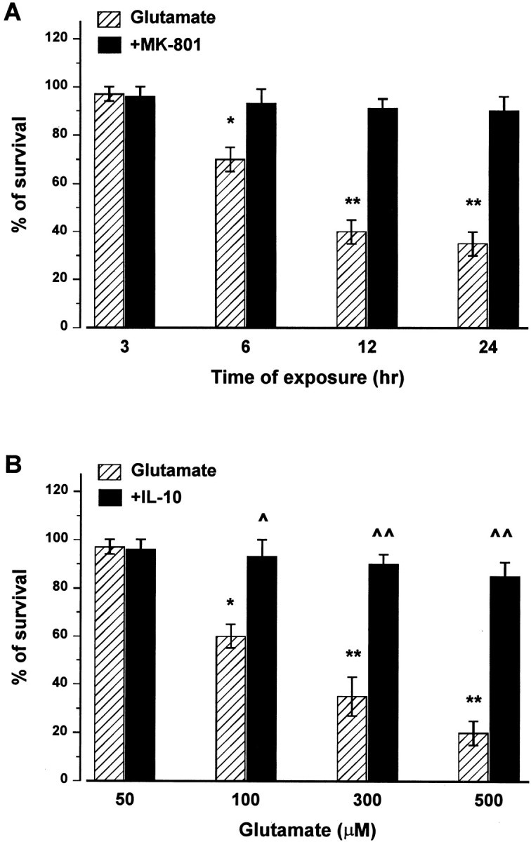
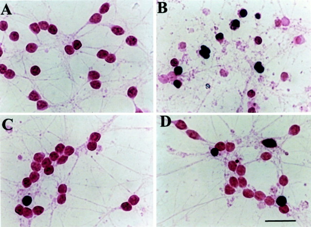
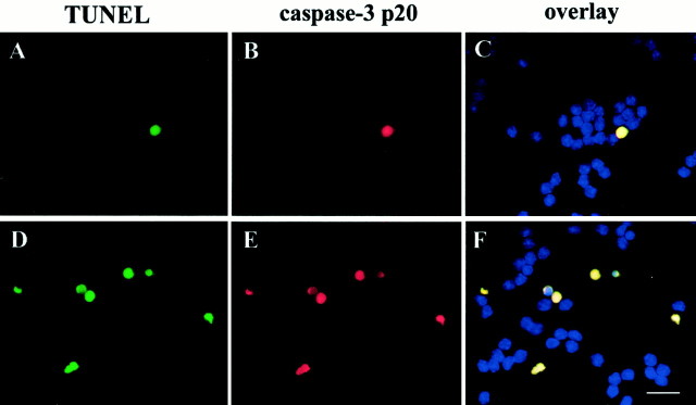
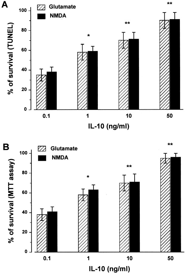
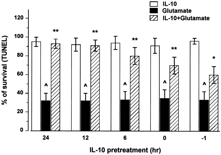
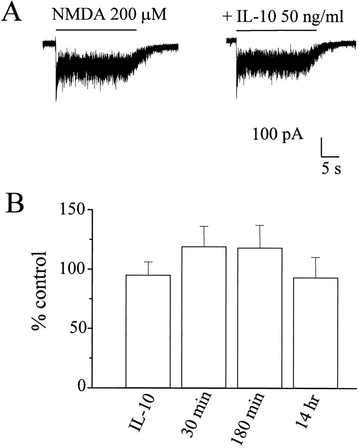
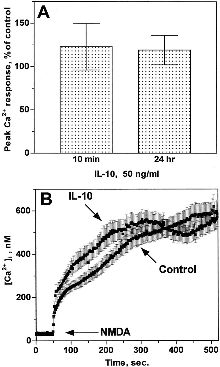
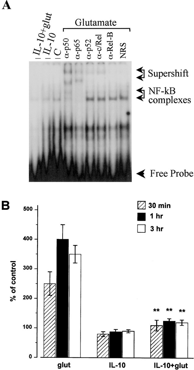
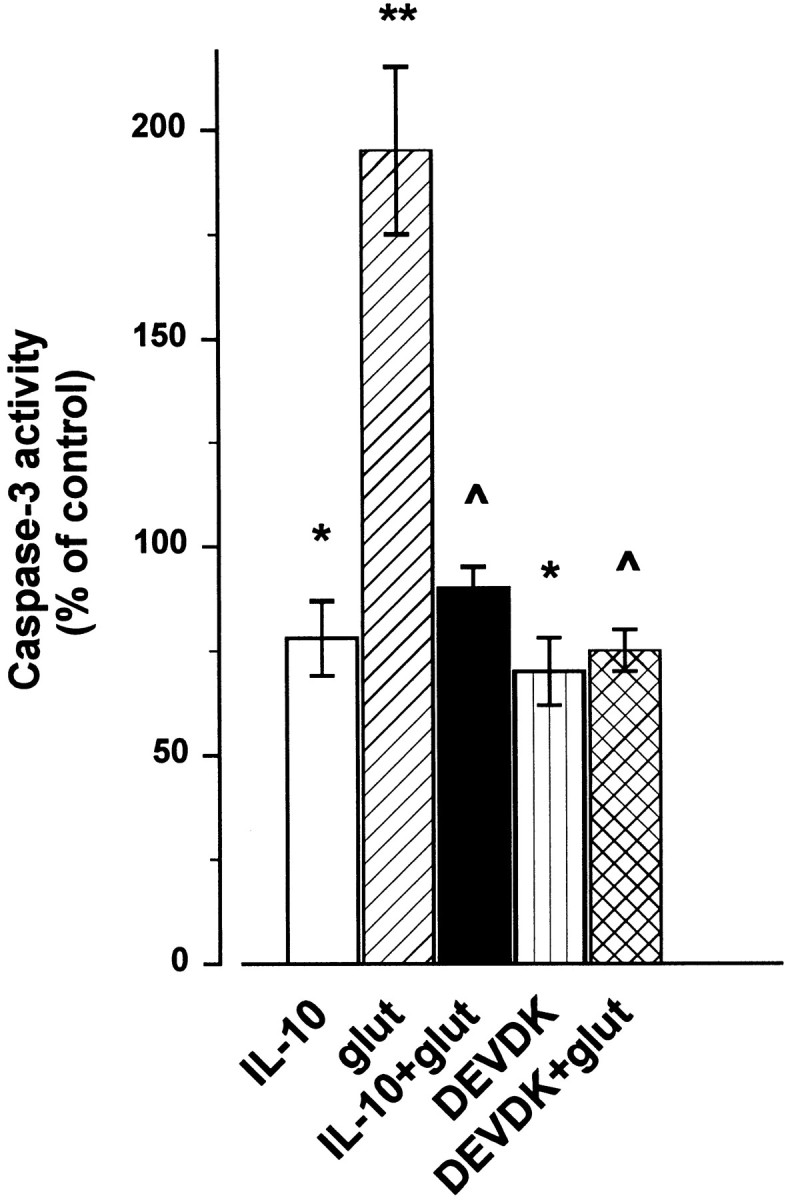
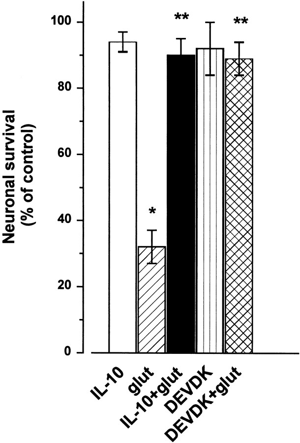
Interleukin-10 (IL-10) has been shown to reduce neuronal degeneration after CNS injury. However, the molecular mechanisms underlying the neuroprotective properties of this cytokine are still under investigation. Glutamate exacerbates secondary injury caused by trauma. Thus, we examined whether IL-10 prevents glutamate-mediated cell death. We used rat cerebellar granule cells in culture because these neurons undergo apoptosis upon exposure to toxic concentrations of glutamate (100-500 microm) or NMDA (300 microm). Pretreatment of cerebellar granule cells with IL-10 (1-50 ng/ml) elicited a dose- and time-dependent reduction of glutamate-induced excitotoxicity. Most importantly, IL-10 reduced the number of apoptotic cells when added to the cultures together or 1 hr after glutamate. Using patch-clamping and fluorescence Ca(2+) imaging techniques, we examined whether IL-10 prevents glutamate toxicity by blocking the function of NMDA channel. IL-10 failed to affect NMDA channel properties and to reduce NMDA-mediated rise in intracellular Ca(2+). Thus, this cytokine appears to prevent glutamate toxicity by a mechanism unrelated to a blockade of NMDA receptor function. Various proteases, such as caspase-3, and transcription factors, such as nuclear factor kappaB (NF-kappaB), have been proposed to participate in glutamate-mediated apoptosis. Thus, we examined whether IL-10 modulates the activity of these apoptotic markers. IL-10 blocked both the glutamate-mediated induction of caspase-3 as well as NF-kappaB DNA binding activity, suggesting that the neuroprotective properties of IL-10 may rely on its ability to block the activity of proapoptotic proteins.


Figures

Fig. 1.
Fig. 1.
Glutamate induces a time- and dose-dependent excitotoxicity blocked by IL-10. A, Cerebellar granule cells were exposed to glutamate (300 μm) in the presence or absence of MK-801 (10 μm) for the indicated times, and then cell viability was measured at the indicated times by MTT assay.B, Neurons were exposed to IL-10 (50 ng/ml) for 14 hr before the addition of different concentrations of glutamate. Cell viability was measured by MTT assay 14 hr after glutamate addition. Data, expressed as percentage of control, are the mean ± SEM of four separate experiments. *p < 0.01, **p < 0.005 versus control; ^p < 0.01, ^^p < 0.005 versus glutamate (ANOVA and Dunnett's test).
Fig. 2.
Fig. 2.
IL-10 reduces the increase of TUNEL-positive cells glutamate mediated. Cerebellar granule cells were exposed to medium alone (A), glutamate (B; 300 μm), or IL-10 (C; 50 ng/ml) for 14 hr, or IL-10 for 14 hr followed by glutamate for 14 hr (D). Cells were then fixed and stained for TUNEL for the determination of apoptosis. In both control and IL-10-treated cultures, 95% of cells were TUNEL-negative, whereas ∼65% of neurons after glutamate treatment were TUNEL-positive (dark brown). Scale bar, 15 μm.
Fig. 3.
Fig. 3.
Caspase-3 immunoreactivity in TUNEL-positive cells. Neurons were exposed to medium alone or glutamate (300 μm) for 3 hr. Determination of apoptotic neurons was performed by TUNEL (A, D) and caspase-3-p20 (B, E) staining.A–C, Control cells; D–F, glutamate-treated cells. Analysis by Magnafire revealed that all TUNEL-positive cells were also positive for caspase-3 (overlay). Scale bar, 15 μm.
Fig. 4.
Fig. 4.
The neuroprotective effect of IL-10 is dose-dependent. Cerebellar granule cells were exposed to different concentrations of IL-10 for 14 hr, and then glutamate or NMDA (300 μm each) was added for additional 14 hr. Cell survival was determined by in situ TUNEL (A) and MTT (B) assay. Data, expressed as percentage of control, are the mean ± SEM of four separate experiments (n = 12 each group). *p < 0.01, **p < 0.005 versus glutamate or NMDA alone (ANOVA and Dunnett's test).
Fig. 5.
Fig. 5.
IL-10 elicits a time-dependent neuroprotection against glutamate. Neurons were exposed to IL-10 alone (50 ng/ml) for 6, 12, and 24 hr before glutamate, to IL-10 and glutamate simultaneously (0), or to glutamate 1 hr before IL-10 (−1). Cell survival was measured 14 hr after glutamate by TUNEL assay. Data are the mean ± SEM of three independent experiments (n = 15 each group). ^p < 0.005 versus control; *p < 0.05, **p < 0.005 versus glutamate (ANOVA and Dunnett's test).
Fig. 6.
Fig. 6.
IL-10 fails to inhibit NMDA-activated currents.A, Current traces from cerebellar granule cells elicited by 200 μm NMDA in the presence and absence of IL-10 (50 ng/ml). NMDA was applied by a Y-tubing device for the duration indicated by the bars. Holding potential, −60 mV.B, Summary of the action of IL-10 treatments in cerebellar granule cells. The left histogram bar labeledIL-10 represents the percentage control of peak currents normalized to the cell capacitance recorded from 31 individual cells during application of NMDA in the presence of IL-10 (50 ng/ml). The other histogram bars represent the percentage control peak current density from at least 10 distinct granule neurons in three distinct sets of experiments in which cells were pretreated with IL-10 (50 ng/ml) for 30 min, 180 min, or 14 hr. Each pointrepresents the mean ± SEM of the ratios of normalized current. No significant differences were found between control and treated cells.
Fig. 7.
Fig. 7.
IL-10 fails to block EAA-mediated [Ca2+]i increase. Ca2+ imaging in cerebellar granule neurons from four independent preparations was performed as described in Materials and Methods. A, Peak [Ca2+]i increase evoked by NMDA in IL-10-treated cells and expressed as percentage of the Ca2+ response in vehicle-treated (control) cells that were imaged in parallel experiments. Data represent mean ± SEM (n = 5) for both 10 min and 24 hr (n = 1 coverslip with 50–99 neurons being imaged simultaneously). B, Ca2+ traces representative of NMDA-evoked [Ca2+]iincrease in neurons pretreated for 24 hr with either vehicle (control) or IL-10. Data (mean ± SEM) represent the [Ca2+]i population mean from 96 (control) and 99 (IL-10) neurons imaged simultaneously.
Fig. 8.
Fig. 8.
IL-10 blocks the glutamate-mediated increase in NF-kB binding activity. Cells were exposed to glutamate (300 μm), IL-10 (50 ng/ml), or a combination of glutamate and IL-10 for various times, nuclear extracts were prepared, and then NF-kB binding activity was measured by EMSA. A, Typical EMSA showing NF-κB complexes (arrows). These complexes were supershifted by α-p50 and p65 but not p52 and c/Rel antibodies. C, Control; glut, glutamate; NRS, normal rabbit serum. B, Relative levels of NF-κB binding activity were quantified by phosphorimager analysis of the band corresponding to NF-κB complexes. Data are the mean ± SEM of three separate experiments, with two to three independent samples each experiment. **p< 0.01 versus glutamate (ANOVA and Dunnett's test).
Fig. 9.
Fig. 9.
IL-10 prevents the increase in caspase-3-like activity evoked by glutamate (glut). Cerebellar granule cells exposed to glutamate (300 μm), IL-10 (50 ng/ml), or DEVDK (100 μm) alone or in combination. IL-10 and DEVDK were added 10 min before glutamate. Caspase-3-like activity was measured 1 hr later. Data are the mean ± SEM of three independent preparations (n = 6 each group). *p < 0.01, **p < 0.005 versus control; ^p < 0.005 versus glutamate (ANOVA and Dunnett's test).
Fig. 10.
Fig. 10.
Inhibition of caspases prevents glutamate-mediated excitotoxicity. Cerebellar granule cells were exposed to glutamate (glut; 300 μm), IL-10 (50 ng/ml), or DEVDK (100 μm) alone or in combination. IL-10 and DEVDK were added 10 min before glutamate. Cell survival was measured 14 hr after by TUNEL assay. Data are the mean ± SEM of three independent preparations (n = 6 each group). *p < 0.005 versus control; **p < 0.005 versus glutamate (ANOVA and Dunnett's test).

References

    1. Anegawa NJ, Lynch DR, Verdoorn TA, Pritchett DB. Transfection of N-methyl-d-aspartate receptors in a nonneuronal cell line leads to cell death. J Neurochem. 1995;64:2004–2012. - PubMed
    1. Ankarcrona M, Dypbukt JM, Bonfoco E, Zhivotovsky B, Orrenius S, Lipton SA, Nicotera P. Glutamate-induced neuronal death: a succession of necrosis or apoptosis depending on mitochondrial function. Neuron. 1995;15:961–973. - PubMed
    1. Baeuerle PA, Baltimore D. NF-κB: ten years after. Cell. 1996;87:13–20. - PubMed
    1. Berkman N, John M, Roesems G, Jose PJ, Barnes PJ, Chung KF. Inhibition of macrophage inflammatory protein-1 α expression by IL-10. J Immunol. 1995;155:4412–4418. - PubMed
    1. Bethea JR, Castro M, Keane RW, Dietrich WD, Yezeirski RP. Traumatic spinal cord injury induces nuclear factor-kB activation. J Neurosci. 1998;18:3251–3260. - PMC - PubMed

Publication types

MeSH terms