Excitatory nicotinic and desensitizing muscarinic (M2) effects on C-nociceptors in isolated rat skin
- PMID: 11312314
- PMCID: PMC6762575
- DOI: 10.1523/JNEUROSCI.21-09-03295.2001
Excitatory nicotinic and desensitizing muscarinic (M2) effects on C-nociceptors in isolated rat skin
Abstract
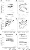
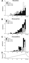
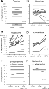
The actions of different cholinergic agonists and antagonists were investigated on nociceptive afferents using the rat skin-saphenous nerve preparation, in vitro. Nicotine was able to weakly excite C-nociceptors and to induce a mild sensitization to heat stimulation (in 77% of tested fibers) in a dose-dependent manner (10(-)6 to 10(-)5 m), but it caused no alteration in mechanical responsiveness tested with von Frey hairs. Muscarine did not induce a significant nociceptor excitation, but almost all fibers exhibited a marked desensitization to mechanical and heat stimuli in a dose-dependent manner (from 10(-)6 to 10(-)4 m). The muscarinic effects could be prevented by the general muscarinic antagonist scopolamine (10(-)5 m), by the M3 antagonist 1,1-dimethyl-4-diphenylacetoxypiperidium oxide (10(-)6 m) co-applied with the M2 antagonist gallamine (10(-)5 m), and by gallamine alone. As positive control we used the relatively M2-selective agonist arecaidine (10(-)6 to 10(-)5 m), obtaining a similar desensitizing effect as with muscarine. Finally, we performed an immunocytochemical study that demonstrated the presence of M2 but not M3 receptors in thin epidermal nerve fibers of the rat hairy skin. Altogether, these data demonstrate opposite effects of nicotinic and muscarinic receptor stimulation on cutaneous nociceptors. M2 receptor-mediated depression of nociceptive responsiveness may convey a therapeutic, i.e., analgesic or antinociceptive, potential.
Figures


References
-
- Bannon AW, Decker MW, Holladay MW, Curzon P, Donnelly-Roberts D, Puttfarcken PS, Bitner RS, Diaz A, Dickenson AH, Porsolt RD, Williams M, Arneric SP. Broad-sprectrum, non-opioid analgesic activity by selective modulation of neuronal nicotinic acetylcholine receptors. Science. 1998;279:77–81. - PubMed
-
- Bauer MB, Murphy S, Gebhart GF. Muscarinic cholinergic stimulation of the nitric oxide-cyclic GMP signaling system in cultured rat sensory neurons. Neuroscience. 1994;62:351–359. - PubMed
-
- Bernardini N, De Stefano ME, Tata AM, Biagioni S, Augusti-Tocco G. Neuronal and non-neuronal cell populations of the avian dorsal root ganglia express muscarinic acetylcholine receptors. Int J Dev Neurosci. 1998;16:365–377. - PubMed
-
- Bernardini N, Levey AI, Augusti-Tocco G. Rat dorsal root ganglia express m1–m4 muscarinic receptor proteins. J Peripher Nerv Syst. 1999;4:222–232. - PubMed
Publication types
MeSH terms
Substances
LinkOut - more resources
Full Text Sources
Research Materials
